File:Gray0465.jpg

Original file (954 × 938 pixels, file size: 207 KB, MIME type: image/jpeg)
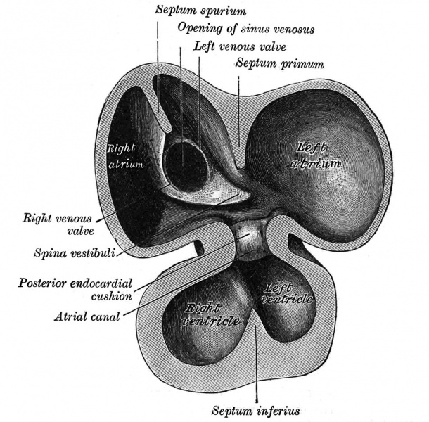
Fig. 465. Interior of dorsal half of heart from a human embryo of about thirty days
From model by Wilhelm His (1831-1904)
The atrial canal is at first a short straight tube connecting the atrial with the ventricular portion of the heart, but its growth is relatively slow, and it becomes overlapped by the atria and ventricles so that its position on the surface of the heart is indicated only by an annular constriction (Fig. 466). Its lumen is reduced to a transverse slit, and two thickenings appear, one on its dorsal and another on its ventral wall. These thickenings, or endocardial cushions (Fig. 465) as they are termed, project into the canal, and, meeting in the middle line, unite to form the septum intermedium which divides the canal into two channels, the future right and left atrioventricular orifices.
The primitive atrium grows rapidly and partially encircles the bulbus cordis; the groove against which the bulbus cordis lies is the first indication of a division into right and left atria. The cavity of the primitive atrium becomes subdivided into right and left chambers by a septum, the septum primum (Fig. 465), which grows downward into the cavity. For a time the atria communicate with each other by an opening, the ostium primum of Bom, below the free margin of the septum. This opening is closed by the union of the septum primum with the septum intermedium, and the communication between the atria is reestablished through an opening which is developed in the upper part of the septum primum; this opening is known as the foramen ovale (ostium secundum of Born) and persists until birth.
| Historic Disclaimer - information about historic embryology pages |
|---|
| Pages where the terms "Historic" (textbooks, papers, people, recommendations) appear on this site, and sections within pages where this disclaimer appears, indicate that the content and scientific understanding are specific to the time of publication. This means that while some scientific descriptions are still accurate, the terminology and interpretation of the developmental mechanisms reflect the understanding at the time of original publication and those of the preceding periods, these terms, interpretations and recommendations may not reflect our current scientific understanding. (More? Embryology History | Historic Embryology Papers) |
- Gray's Images: Development | Lymphatic | Neural | Vision | Hearing | Somatosensory | Integumentary | Respiratory | Gastrointestinal | Urogenital | Endocrine | Surface Anatomy | iBook | Historic Disclaimer
| Historic Disclaimer - information about historic embryology pages |
|---|
| Pages where the terms "Historic" (textbooks, papers, people, recommendations) appear on this site, and sections within pages where this disclaimer appears, indicate that the content and scientific understanding are specific to the time of publication. This means that while some scientific descriptions are still accurate, the terminology and interpretation of the developmental mechanisms reflect the understanding at the time of original publication and those of the preceding periods, these terms, interpretations and recommendations may not reflect our current scientific understanding. (More? Embryology History | Historic Embryology Papers) |
| iBook - Gray's Embryology | |
|---|---|

|
|
Reference
Gray H. Anatomy of the human body. (1918) Philadelphia: Lea & Febiger.
Cite this page: Hill, M.A. (2024, April 27) Embryology Gray0465.jpg. Retrieved from https://embryology.med.unsw.edu.au/embryology/index.php/File:Gray0465.jpg
- © Dr Mark Hill 2024, UNSW Embryology ISBN: 978 0 7334 2609 4 - UNSW CRICOS Provider Code No. 00098G
File history
Click on a date/time to view the file as it appeared at that time.
| Date/Time | Thumbnail | Dimensions | User | Comment | |
|---|---|---|---|---|---|
| current | 19:21, 23 August 2014 |  | 954 × 938 (207 KB) | Z8600021 (talk | contribs) | |
| 19:18, 23 August 2014 |  | 1,276 × 1,000 (228 KB) | Z8600021 (talk | contribs) | {{Historic Disclaimer}} {{Gray Anatomy}} Category:Cardiovascular Category:Blood Vessel[Category:Chicken]] Category:Historic Embryology Category:Gray's 1918 Anatomy |
You cannot overwrite this file.
File usage
The following page uses this file:
