File:Gray0464.jpg

Original file (993 × 961 pixels, file size: 258 KB, MIME type: image/jpeg)
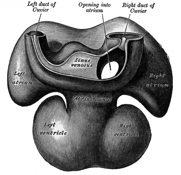
Fig. 464. Dorsal surface of heart of human embryo of thirty-five days
From model by Wilhelm His (1831-1904)
The sinus venosus is at first situated in the septum transversum (a layer of mesoderm in which the liver and the central tendon of the diaphragm are developed) behind the primitive atrium, and is formed by the union of the vitelline veins. The veins or ducts of Cuvier from the body of the embryo and the umbilical veins from the placenta subsequently open into it (Fig. 463) . The sinus is at first place transversely, and opens by a median aperture into the primitive atrium. Soon, however, it assumes an oblique position, and becomes crescentic in form; its right half or horn increases more rapidly than the left, and the opening into the atrium now communicates with the right portion of the atrial cavity. The right horn and transverse portion of the sinus ultimately become incorporated with and form a part of the adult right atrium, the line of union between it and the auricula being indicated in the interior of the atrium by a vertical crest, the crista terminalis of His. The left horn, which ultimately receives only the left duct of Cuvier, persists as the coronary sinus (Fig. 464) . The vitelline and umbilical veins are soon replaced by a single vessel, the inferior vena cava, and the three veins (inferior vena cava and right and left Cuvierian ducts) open into the dorsal aspect of the atrium by a common slit-like aperture (Fig. 465). The upper part of this aperture represents the opening of the permanent superior vena cava, the lower that of the inferior vena cava, and the intermediate part the orifice of the coronary sinus. The slit-like aperture lies obliquely, and is guarded by two halves, the right and left venous valves; above the opening these unite with each other and are continuous with a fold named the septum spurium; below the opening they fuse to form a triangular thickening — the spina vestibuli. The right venous valve is retained; a small septum, the sinus septum, grows from the posterior wall of the sinus venosus to fuse 'with the valve and divide it into two parts — an upper, the valve of the inferior vena cava, and a lower, the valve of the coronary sinus (Fig. 468). The extreme upper portion of the right venous valve, together with the septum spurium, form the crista terminalis already mentioned. The upper and middle thirds of the left venous valve disappear; the lower third is continued into the spina vestibuli, md later fuses with the septum secundum of the atria and takes part in the formation of the limbus fossae ovalis.
| Historic Disclaimer - information about historic embryology pages |
|---|
| Pages where the terms "Historic" (textbooks, papers, people, recommendations) appear on this site, and sections within pages where this disclaimer appears, indicate that the content and scientific understanding are specific to the time of publication. This means that while some scientific descriptions are still accurate, the terminology and interpretation of the developmental mechanisms reflect the understanding at the time of original publication and those of the preceding periods, these terms, interpretations and recommendations may not reflect our current scientific understanding. (More? Embryology History | Historic Embryology Papers) |
- Gray's Images: Development | Lymphatic | Neural | Vision | Hearing | Somatosensory | Integumentary | Respiratory | Gastrointestinal | Urogenital | Endocrine | Surface Anatomy | iBook | Historic Disclaimer
| Historic Disclaimer - information about historic embryology pages |
|---|
| Pages where the terms "Historic" (textbooks, papers, people, recommendations) appear on this site, and sections within pages where this disclaimer appears, indicate that the content and scientific understanding are specific to the time of publication. This means that while some scientific descriptions are still accurate, the terminology and interpretation of the developmental mechanisms reflect the understanding at the time of original publication and those of the preceding periods, these terms, interpretations and recommendations may not reflect our current scientific understanding. (More? Embryology History | Historic Embryology Papers) |
| iBook - Gray's Embryology | |
|---|---|

|
|
Reference
Gray H. Anatomy of the human body. (1918) Philadelphia: Lea & Febiger.
Cite this page: Hill, M.A. (2024, April 27) Embryology Gray0464.jpg. Retrieved from https://embryology.med.unsw.edu.au/embryology/index.php/File:Gray0464.jpg
- © Dr Mark Hill 2024, UNSW Embryology ISBN: 978 0 7334 2609 4 - UNSW CRICOS Provider Code No. 00098G
File history
Click on a date/time to view the file as it appeared at that time.
| Date/Time | Thumbnail | Dimensions | User | Comment | |
|---|---|---|---|---|---|
| current | 19:21, 23 August 2014 |  | 993 × 961 (258 KB) | Z8600021 (talk | contribs) | |
| 19:19, 23 August 2014 |  | 1,238 × 1,000 (276 KB) | Z8600021 (talk | contribs) | {{Historic Disclaimer}} {{Gray Anatomy}} Category:Cardiovascular Category:Blood Vessel[Category:Chicken]] Category:Historic Embryology Category:Gray's 1918 Anatomy |
You cannot overwrite this file.
File usage
The following page uses this file:
