File:Gray0193.jpg

Original file (719 × 1,057 pixels, file size: 293 KB, MIME type: image/jpeg)
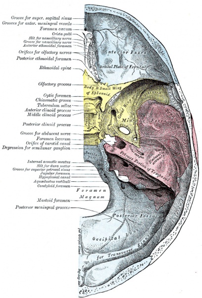
Fig. 193. Upper Surface of the Base of the Skull
The upper surface of the base of the skull or floor of the cranial cavity presents three fossæ, called the anterior, middle, and posterior cranial fuss.
Anterior Fossa
(fossa cranii anterior) The floor of the anterior fossa is formed by the orbital plates of the frontal, the cribriform plate of the ethmoid, and the small wings and front part of the body of the sphenoid; it is limited behind by the posterior borders of the small wings of the sphenoid and by the anterior margin of the chiasmatic groove. It is traversed by the frontoethmoidal, sphenoethmoidal, and sphenofrontal sutures. Its lateral portions roof in the orbital cavities and support the frontal lobes of the cerebrum; they are convex and marked by depressions for the brain convolutions, and grooves for branches of the meningeal vessels. The central portion corresponds with the roof of the nasal cavity, and is markedly depressed on either side of the crista galli. It presents, in and near the median line, from before backward, the commencement of the frontal crest for the attachment of the falx cerebri; the foramen cecum, between the frontal bone and the crista galli of the ethmoid, which usually transmits a small vein from the nasal cavity to the superior sagittal sinus; behind the foramen cecum, the crista galli, the free margin of which affords attachment to the falx cerebri; on either side of the crista galli, the olfactory groove formed by the cribriform plate, which supports the olfactory bulb and presents foramina for the transmission of the olfactory nerves, and in front a slit-like opening for the nasociliary nerve. Lateral to either olfactory groove are the internal openings of the anterior and posterior ethmoidal foramina; the anterior, situated about the middle of the lateral margin of the olfactory groove, transmits the anterior ethmoidal vessels and the nasociliary nerve; the nerve runs in a groove along the lateral edge of the cribriform plate to the slit-like opening above mentioned; the posterior ethmoidal foramen opens at the back part of this margin under cover of the projecting lamina of the sphenoid, and transmits the posterior ethmoidal vessels and nerve. Farther back in the middle line is the ethmoidal spine, bounded behind by a slight elevation separating two shallow longitudinal grooves which support the olfactory lobes. Behind this is the anterior margin of the chiasmatic groove, running lateralward on either side to the upper margin of the optic foramen.
Middle Fossa
(fossa cranii media) The middle fossa, deeper than the preceding, is narrow in the middle, and wide at the sides of the skull. It is bounded in front by the posterior margins of the small wings of the sphenoid, the anterior clinoid processes, and the ridge forming the anterior margin of the chiasmatic groove; behind, by the superior angles of the petrous portions of the temporals and the dorsum sellæ; laterally by the temporal squamæ, sphenoidal angles of the parietals, and great wings of the sphenoid. It is traversed by the squamosal, sphenoparietal, sphenosquamosal, and sphenopetrosal sutures.
The middle part of the fossa presents, in front, the chiasmatic groove and tuberculum sellæ; the chiasmatic groove ends on either side at the optic foramen, which transmits the optic nerve and ophthalmic artery to the orbital cavity. Behind the optic foramen the anterior clinoid process is directed backward and medialward and gives attachment to the tentorium cerebelli. Behind the tuberculum sellæ is a deep depression, the sella turcica, containing the fossa hypophyseos, which lodges the hypophysis, and presents on its anterior wall the middle clinoid processes. The sella turcica is bounded posteriorly by a quadrilateral plate of bone, the dorsum sellæ, the upper angles of which are surmounted by the posterior clinoid processes: these afford attachment to the tentorium cerebelli, and below each is a notch for the abducent nerve. On either side of the sella turcica is the carotid groove, which is broad, shallow, and curved somewhat like the italic letter f. It begins behind at the foramen lacerum, and ends on the medial side of the anterior clinoid process, where it is sometimes converted into a foramen (carotico-clinoid) by the union of the anterior with the middle clinoid process; posteriorly, it is bounded laterally by the lingula. This groove lodges the cavernous sinus and the internal carotid artery, the latter being surrounded by a plexus of sympathetic nerves.
The lateral parts of the middle fossa are of considerable depth, and support the temporal lobes of the brain. They are marked by depressions for the brain convolutions and traversed by furrows for the anterior and posterior branches of the middle meningeal vessels. These furrows begin near the foramen spinosum, and the anterior runs forward and upward to the sphenoidal angle of the parietal, where it is sometimes converted into a bony canal; the posterior runs lateralward and backward across the temporal squama and passes on to the parietal near the middle of its lower border. The following apertures are also to be seen. In front is the superior orbital fissure, bounded above by the small wing, below, by the great wing, and medially, by the body of the sphenoid; it is usually completed laterally by the orbital plate of the frontal bone. It transmits to the orbital cavity the oculomotor, the trochlear, the ophthalmic division of the trigeminal, and the abducent nerves, some filaments from the cavernous plexus of the sympathetic, and the orbital branch of the middle meningeal artery; and from the orbital cavity a recurrent branch from the lacrimal artery to the dura mater, and the ophthalmic veins. Behind the medial end of the superior orbital fissure is the foramen rotundum, for the passage of the maxillary nerve. Behind and lateral to the foramen rotundum is the foramen ovale, which transmits the mandibular nerve, the accessory meningeal artery, and the lesser superficial petrosal nerve. 50 Medial to the foramen ovale is the foramen Vesalii, which varies in size in different individuals, and is often absent; when present, it opens below at the lateral side of the scaphoid fossa, and transmits a small vein. Lateral to the foramen ovale is the foramen spinosum, for the passage of the middle meningeal vessels, and a recurrent branch from the mandibular nerve. Medial to the foramen ovale is the foramen lacerum; in the fresh state the lower part of this aperture is filled up by a layer of fibrocartilage, while its upper and inner parts transmit the internal carotid artery surrounded by a plexus of sympathetic nerves. The nerve of the pterygoid canal and a meningeal branch from the ascending pharyngeal artery pierce the layer of fibrocartilage. On the anterior surface of the petrous portion of the temporal bone are seen the eminence caused by the projection of the superior semicircular canal; in front of and a little lateral to this a depression corresponding to the roof of the tympanic cavity; the groove leading to the hiatus of the facial canal, for the transmission of the greater superficial petrosal nerve and the petrosal branch of the middle meningeal artery; beneath it, the smaller groove, for the passage of the lesser superficial petrosal nerve; and, near the apex of the bone, the depression for the semilunar ganglion and the orifice of the carotid canal.
The Posterior Fossa
(fossa cranii posterior) The posterior fossa is the largest and deepest of the three. It is formed by the dorsum sellæ and clivus of the sphenoid, the occipital, the petrous and mastoid portions of the temporals, and the mastoid angles of the parietal bones; it is crossed by the occipitomastoid and the parietomastoid sutures, and lodges the cerebellum, pons, and medulla oblongata. It is separated from the middle fossa in and near the median line by the dorsum sellæ of the sphenoid and on either side by the superior angle of the petrous portion of the temporal bone. This angle gives attachment to the tentorum cerebelli, is grooved for the superior petrosal sinus, and presents at its medial end a notch upon which the trigeminal nerve rests. The fossa is limited behind by the grooves for the transverse sinuses. In its center is the foramen magnum, on either side of which is a rough tubercle for the attachment of the alar ligaments; a little above this tubercle is the canal, which transmits the hypoglossal nerve and a meningeal branch from the ascending pharyngeal artery. In front of the foramen magnum the basilar portion of the occipital and the posterior part of the body of the sphenoid form a grooved surface which supports the medulla oblongata and pons; in the young skull these bones are joined by a synchondrosis. This grooved surface is separated on either side from the petrous portion of the temporal by the petro-occipital fissure, which is occupied in the fresh state by a plate of cartilage; the fissure is continuous behind with the jugular foramen, and its margins are grooved for the inferior petrosal sinus. The jugular foramen is situated between the lateral part of the occipital and the petrous part of the temporal. The anterior portion of this foramen transmits the inferior petrosal sinus; the posterior portion, the transverse sinus and some meningeal branches from the occipital and ascending pharyngeal arteries; and the intermediate portion, the glossopharyngeal, vagus, and accessory nerves. Above the jugular foramen is the internal acoustic meatus, for the facial and acoustic nerves and internal auditory artery; behind and lateral to this is the slit-like opening leading into the aquæductus vestibuli, which lodges the ductus endolymphaticus; while between these, and near the superior angle of the petrous portion, is a small triangular depression, the remains of the fossa subarcuata, which lodges a process of the dura mater and occasionally transmits a small vein. Behind the foramen magnum are the inferior occipital fossæ, which support the hemispheres of the cerebellum, separated from one another by the internal occipital crest, which serves for the attachment of the falx cerebelli, and lodges the occipital sinus. The posterior fossæ are surmounted by the deep grooves for the transverse sinuses. Each of these channels, in its passage to the jugular foramen, grooves the occipital, the mastoid angle of the parietal, the mastoid portion of the temporal, and the jugular process of the occipital, and ends at the back part of the jugular foramen. Where this sinus grooves the mastoid portion of the temporal, the orifice of the mastoid foramen may be seen; and, just previous to its termination, the condyloid canal opens into it; neither opening is constant.
- Gray's Images: Development | Lymphatic | Neural | Vision | Hearing | Somatosensory | Integumentary | Respiratory | Gastrointestinal | Urogenital | Endocrine | Surface Anatomy | iBook | Historic Disclaimer
| Historic Disclaimer - information about historic embryology pages |
|---|
 |
| iBook - Gray's Embryology | |
|---|---|

|
|
Reference
Gray H. Anatomy of the human body. (1918) Philadelphia: Lea & Febiger.
Cite this page: Hill, M.A. (2024, April 27) Embryology Gray0193.jpg. Retrieved from https://embryology.med.unsw.edu.au/embryology/index.php/File:Gray0193.jpg
- © Dr Mark Hill 2024, UNSW Embryology ISBN: 978 0 7334 2609 4 - UNSW CRICOS Provider Code No. 00098G
File history
Click on a date/time to view the file as it appeared at that time.
| Date/Time | Thumbnail | Dimensions | User | Comment | |
|---|---|---|---|---|---|
| current | 11:08, 5 November 2015 | | 719 × 1,057 (293 KB) | Z8600021 (talk | contribs) |
You cannot overwrite this file.
File usage
The following page uses this file: