File:Gray0130.jpg
Gray0130.jpg (535 × 600 pixels, file size: 95 KB, MIME type: image/jpeg)
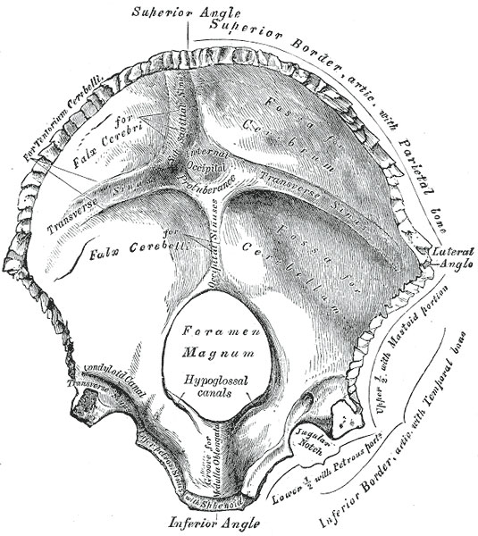
Fig. 130. Human Occipital Bone
The occipital bone (Figs. 129, 130), situated at the back and lower part of the cranium, is trapezoid in shape and curved on itself. It is pierced by a large oval aperture, the foramen magnum, through which the cranial cavity communicates with the vertebral canal.
The curved, expanded plate behind the foramen magnum is named the squama; the thick, somewhat quadrilateral piece in front of the foramen is called the basilar part, whilst on either side of the foramen is the lateral portion.
The internal surface is deeply concave and divided into four fossæ by a cruciate eminence. The upper two fossæ are triangular and lodge the occipital lobes of the cerebrum; the lower two are quadrilateral and accommodate the hemispheres of the cerebellum. At the point of intersection of the four divisions of the cruciate eminence is the internal occipital protuberance. From this protuberance the upper division of the cruciate eminence runs to the superior angle of the bone, and on one side of it (generally the right) is a deep groove, the sagittal sulcus, which lodges the hinder part of the superior sagittal sinus; to the margins of this sulcus the falx cerebri is attached. The lower division of the cruciate eminence is prominent, and is named the internal occipital crest; it bifurcates near the foramen magnum and gives attachment to the falx cerebelli; in the attached margin of this falx is the occipital sinus, which is sometimes duplicated. In the upper part of the internal occipital crest, a small depression is sometimes distinguishable; it is termed the vermian fossa since it is occupied by part of the vermis of the cerebellum. Transverse grooves, one on either side, extend from the internal occipital protuberance to the lateral angles of the bone; those grooves accommodate the transverse sinuses, and their prominent margins give attachment to the tentorium cerebelli. The groove on the right side is usually larger than that on the left, and is continuous with that for the superior sagittal sinus. Exceptions to this condition are, however, not infrequent; the left may be larger than the right or the two may be almost equal in size. The angle of union of the superior sagittal and transverse sinuses is named the confluence of the sinuses (torcular Herophili 25), and its position is indicated by a depression situated on one or other side of the protuberance.
- Gray's Images: Development | Lymphatic | Neural | Vision | Hearing | Somatosensory | Integumentary | Respiratory | Gastrointestinal | Urogenital | Endocrine | Surface Anatomy | iBook | Historic Disclaimer
| Historic Disclaimer - information about historic embryology pages |
|---|
| Pages where the terms "Historic" (textbooks, papers, people, recommendations) appear on this site, and sections within pages where this disclaimer appears, indicate that the content and scientific understanding are specific to the time of publication. This means that while some scientific descriptions are still accurate, the terminology and interpretation of the developmental mechanisms reflect the understanding at the time of original publication and those of the preceding periods, these terms, interpretations and recommendations may not reflect our current scientific understanding. (More? Embryology History | Historic Embryology Papers) |
| iBook - Gray's Embryology | |
|---|---|

|
|
Reference
Gray H. Anatomy of the human body. (1918) Philadelphia: Lea & Febiger.
Cite this page: Hill, M.A. (2024, April 27) Embryology Gray0130.jpg. Retrieved from https://embryology.med.unsw.edu.au/embryology/index.php/File:Gray0130.jpg
- © Dr Mark Hill 2024, UNSW Embryology ISBN: 978 0 7334 2609 4 - UNSW CRICOS Provider Code No. 00098G
File history
Click on a date/time to view the file as it appeared at that time.
| Date/Time | Thumbnail | Dimensions | User | Comment | |
|---|---|---|---|---|---|
| current | 11:06, 5 November 2015 |  | 535 × 600 (95 KB) | Z8600021 (talk | contribs) | ==Fig. 130. Human occipital bone== Inner surface. |
You cannot overwrite this file.
File usage
The following page uses this file:

