File:GladstoneWakeley1937 plate06.jpg
Original file (1,688 × 2,521 pixels, file size: 556 KB, MIME type: image/jpeg)
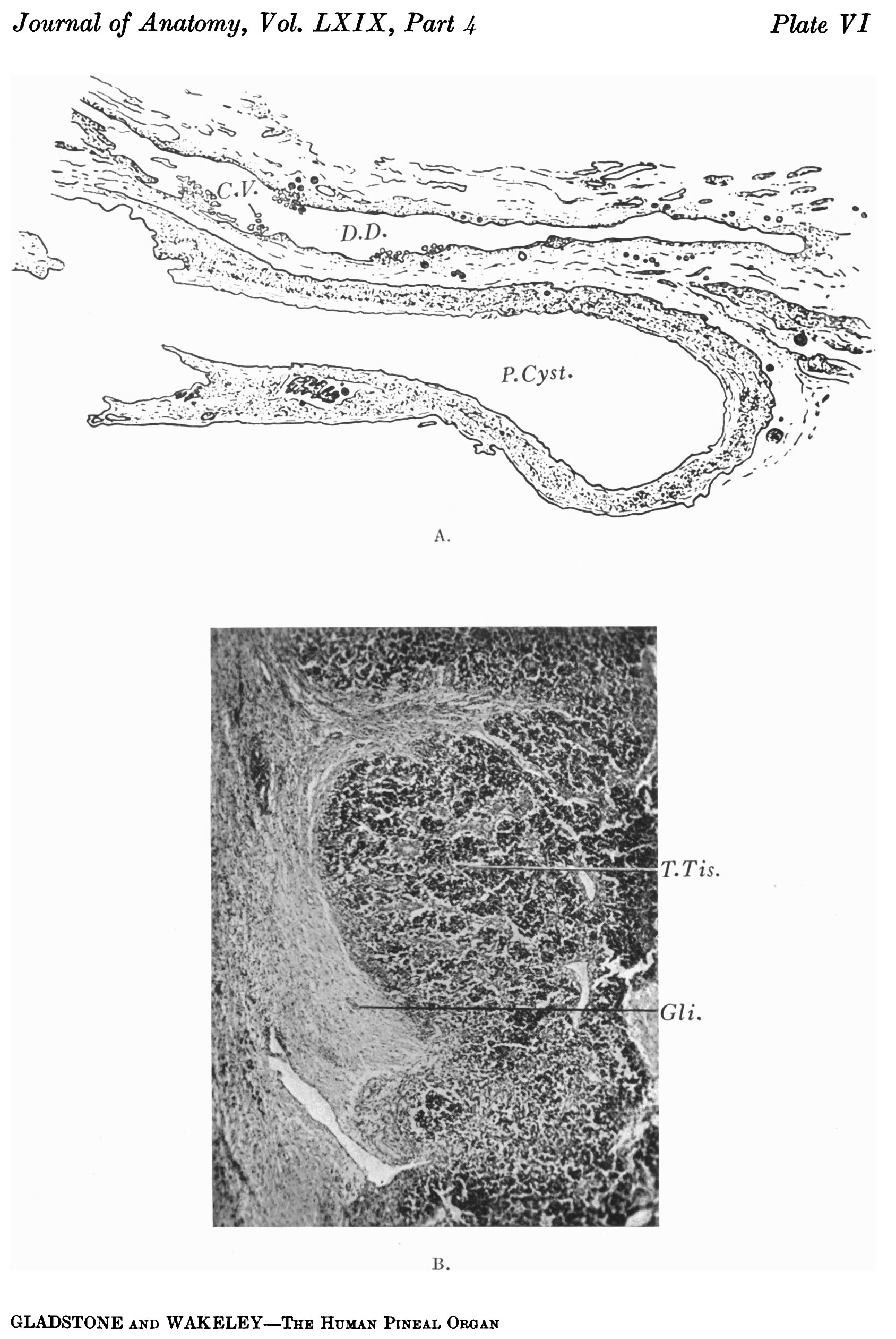
Plate VI.
A. Section of pineal cyst shown in Plate V C and E. The drawing also shows the relation of the dorsal diverticulum to the pineal gland, and chorioidal villi projecting into its lumen. Corpora arenacea are present in the tissue surrounding the pineal cyst and the diverticulum, and are also imbedded in their walls.
B. Section of pineal tumour showing the lobulated arrangement of the tumour tissue, and tracts of degenerating glial tissue, containing thin-walled blood vessels.
Reference
Gladstone RJ. and Wakeley CPG. Development and histogenesis of the human pineal organ. (1937) J. Anat., 19(4): 431-454.
Cite this page: Hill, M.A. (2024, May 1) Embryology GladstoneWakeley1937 plate06.jpg. Retrieved from https://embryology.med.unsw.edu.au/embryology/index.php/File:GladstoneWakeley1937_plate06.jpg
- © Dr Mark Hill 2024, UNSW Embryology ISBN: 978 0 7334 2609 4 - UNSW CRICOS Provider Code No. 00098G
File history
Click on a date/time to view the file as it appeared at that time.
| Date/Time | Thumbnail | Dimensions | User | Comment | |
|---|---|---|---|---|---|
| current | 10:17, 4 January 2017 | | 1,688 × 2,521 (556 KB) | Z8600021 (talk | contribs) | ==Plate VI.== ===Reference=== {{Ref-GladstoneWakeley1937}} {{Footer}} |
You cannot overwrite this file.
File usage
The following page uses this file: