File:GladstoneWakeley1937 plate03.jpg
Original file (1,827 × 2,504 pixels, file size: 381 KB, MIME type: image/jpeg)
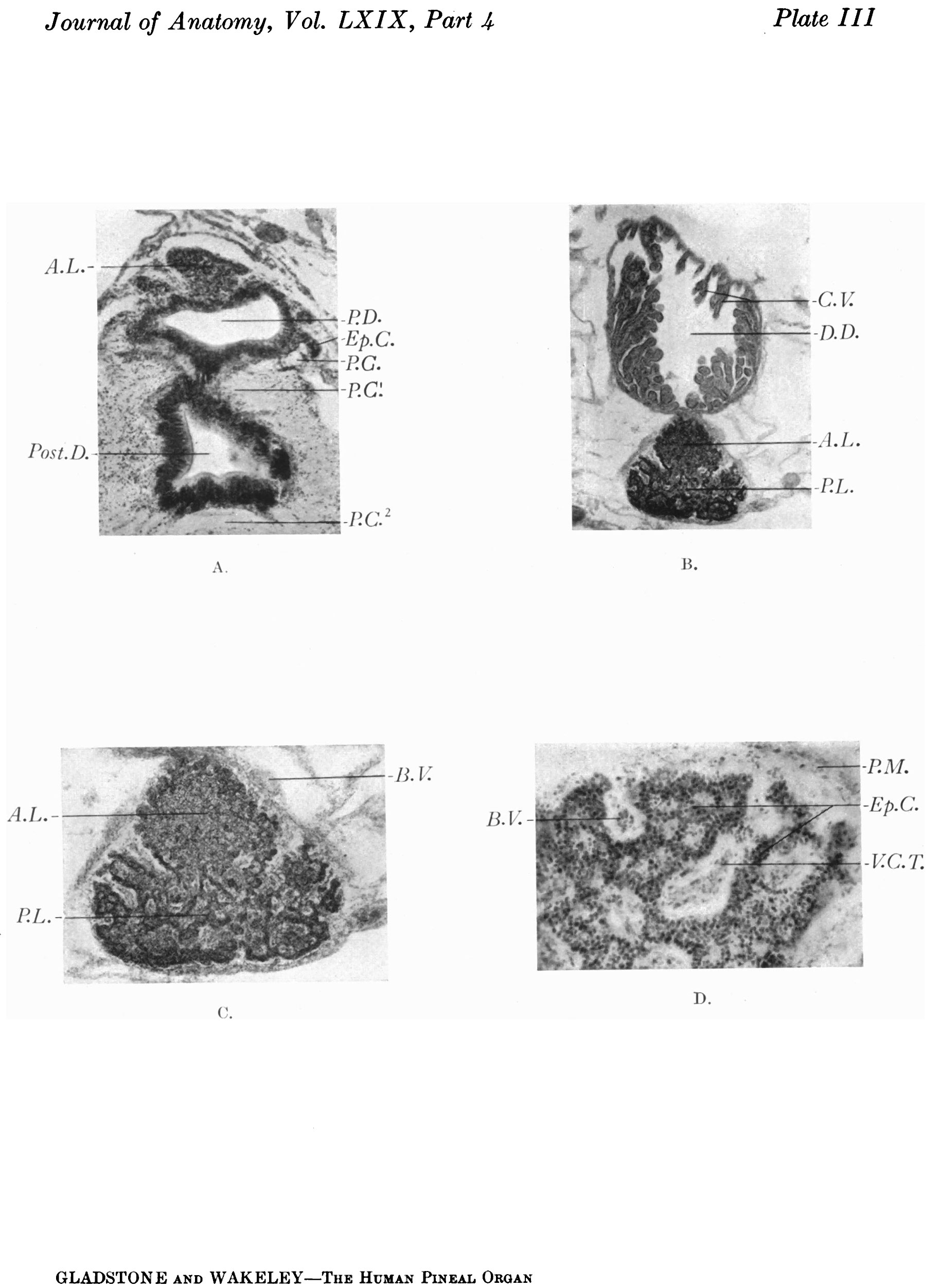
Plate III
Transverse sections of the pineal region of a 6-cm. human embryo, and of a 4.5 months’ foetus.
A. Section through the basal part of the main pineal diverticulum of a 6-cm. human embryo showing, in the upper part of the photograph, the solid anterior lobe. Below this is the main pineal diverticulum, the wall of which shows proliferating cords of ependymal cells growing outward, into the surrounding tissue. Below the pineal evagination is a section through the infrapineal recess, the epithelial lining of which is assuming a columnar type. Fibres of the posterior commissure are seen at the sides and below the pineal region.
B. Coronal section through the pineal region of a 4} months’ human foetus showing the relation of the dorsal diverticulum (suprapineal recess) to the pineal gland. Clusters of elongated chorioidal villi project into the lumen of the diverticulum. The pineal gland shows partial subdivision into an anterior and posterior lobe.
C. The pineal gland more highly magnified showing the ingrowth of vascular processes of the pia mater between the outgrowing neuro-epithelial cords.
D. Peripheral portion of the gland x55 D., showing pale areas containing a central core of vascular pia mater, alternating with dark zones, composed of neuro-epithelial cords.
| Figure Abbreviations |
|---|
| A.B.D. Anterior bilobed diverticulum.
Ant.D. Anterior diverticulum. A.L. Anterior lobe. Aq.C. Aqueductus cerebri. B. V. Blood vessel. C'. Constriction between “pineal sac” and “pineal eye”. Ca. Capsule. Cap. Capillary. Cav. Cavity. Cbl. Cerebellum. C.C. Corpus callosum. C'.E., C.Ep. Columuar epithelium. C.H. Cerebral hemisphere. Ch.P. Chorioid plexus. C'h.P.V. III Chorioid plexus of third ventricle. C'h.P.L. V. Chorioid plexus of lateral ventricle. C.M. Corpus mammillaris. C.N. III Cranial nerve III. C.N. IV Cranial nerve IV. 0.N. V Cranial nerve V. Cp. Capillary. C.T. Connective tissue. C.T.C’. Connective tissue cells. Cr.C'. Cranial capsule. Cyst. Cyst containing chorioidal villi. C. V. Chorioidal villi. D.D., D.D.’, D.D.” Dorsal diverticulum and its subdivisions. D.S. Dorsal sac. D.M. Dura mater. E.L.M. External limiting membrane. End.S. Endothelial space. Ep. Ependyma. Ep.D. Ependymal diverticulum (dorsal sac). Epd. Epidermis. Ep.Z. Ependymal zone. Ep.C. Epithelial column. Iv'.(.'. Vessels in fibrous capsule. F .F’. F ornix. G.C. V. Great cerebral vein (Galen). G'li., G.S'l2. Glial sheath. Gl.St. Glial stratum (pseudo-epithelium). H.G. Habenular ganglion. I.S.S. Inferior sagittal sinus. I.L.S. Interlobar septum. I.Lr.S. Interlobular septum. Inf. Infundibulum. I.P.R. Infrapineal recess. L. Lobule. Le. Lens. L.N.F.I’.S. Layer of nerve fibres of pineal sac. L.N.F.R. Layer of nerve fibres of retina. Lmn. Lumen. M.B. Meynert’s bundle. M.Z. Marginal zone. N.Z. Nuclear zone. 0p.P.St. Opening of pineal stalk. 0.T. Optic thalamus. P. Pulvinar. Pa. ? Paraphysis. 'Pa.C. Parenchyma cell. Pa.0. Parietal organ (pineal eye). Par. Parenchyma. P.B. Pineal Body. P.O. Pineal cells. P.Co., P.C'., P.0.1, P.C.3 Posterior commissure. P.Cyst. Pineal cyst. P.D. Pineal diverticulum. P.E. Pineal eye (parietal organ). P.I.P. Posterior-intercalary plate. P.L. Posterior lobe. P.M., P.M.1, P.M.’ Pia mater. P.M.D. Posterior median diverticulum. P.0. Pineal organ. Post.D. Posterior diverticulum. P.P.0. Peduncle of pineal organ. P.R. Pineal recess. Pr. Ep. Epithelial process. P.S. Pineal sac. P.Sh. Pial sheath. P.St. Pineal stalk. P. V. Pons Varolii. P. V.A . Post-velar arch. Pv.Sp. Perivascular space. Q.P. Quadrigeminal plate. R. Retina. Ros. “ Rosettes ” . R.P. Rathke’s pouch. S. Septum interhemisphericum. S.C’. Superior commissure (habenular commissure). S.C'.0. Subcommissural organ. S.Co. Sinus confluens. S.0ol. Superior colliculus. S.D. Secondary diverticulum. S.E. condaly evagination. S.D.N. Small darkly stained nucleus. Sp. Space. S.P.L. Splenium. S.T. Sinus transversus. SLO. Stieda’s organ (terminal vesicle). Sub.ep. Subependymal layer. T. Tongue. T.Cbl. Tentorium cerebelli. Th. Thalamus. Tag. Tegmentum. Thr. Thrombus. V.O'.M. Great cerebral vein (V.M. Galeni). V.C.T. Vascular connective tissue. V.F. Velar fold. V.M. Ventriculus mesencephalicus. V.L. Ventriculus lateralis. V. III Third ventricle. V. IV Fourth ventricle. V.S. Venous sinuses. |
| Historic Disclaimer - information about historic embryology pages |
|---|
 |
- Links: fig 1 | fig 2 | fig 3 | fig 4 | fig 5 | fig 6 | fig 7 | fig 8 | fig 9 | fig 10 | fig 18 | plate 1 | plate 2 | plate 3 | plate 4 | plate 5 | plate 6 | 1937 Gladstone Wakeley | Endocrine - Pineal Development | Category:Pineal
Reference
Gladstone RJ. and Wakeley CPG. Development and histogenesis of the human pineal organ. (1937) J. Anat., 19(4): 431-454.
Cite this page: Hill, M.A. (2024, April 27) Embryology GladstoneWakeley1937 plate03.jpg. Retrieved from https://embryology.med.unsw.edu.au/embryology/index.php/File:GladstoneWakeley1937_plate03.jpg
- © Dr Mark Hill 2024, UNSW Embryology ISBN: 978 0 7334 2609 4 - UNSW CRICOS Provider Code No. 00098G
File history
Click on a date/time to view the file as it appeared at that time.
| Date/Time | Thumbnail | Dimensions | User | Comment | |
|---|---|---|---|---|---|
| current | 10:28, 4 January 2017 | | 1,827 × 2,504 (381 KB) | Z8600021 (talk | contribs) |
You cannot overwrite this file.
File usage
The following page uses this file: