File:GladstoneHamilton1941 text-fig10.jpg

Original file (1,149 × 1,493 pixels, file size: 364 KB, MIME type: image/jpeg)
Text-fig. 10. The photograph shows the junction of the connecting stalk with the chorionic mesoderm
And the advanced stage of development of the vessels in the stalk, adjacent parietal mesenchyme, and mesenchymal tissue of the In the cavity of the chorionic vesicle isolated groups of degenerate mesenchymal cells and free nuclei are visible. The connexions of the villi with the decidua, and of the intervillous spaces with the basal sinus, are also clearly shown. Section no. 144. x 100.
Reference
Gladstone RJ. and Hamilton WJ. A presomite human embryo (Shaw) with primitive streak and chorda canal with special reference to the development of the vascular system. (1941) Amer. J Anat. 76(1): 9-44.
Cite this page: Hill, M.A. (2024, April 27) Embryology GladstoneHamilton1941 text-fig10.jpg. Retrieved from https://embryology.med.unsw.edu.au/embryology/index.php/File:GladstoneHamilton1941_text-fig10.jpg
- © Dr Mark Hill 2024, UNSW Embryology ISBN: 978 0 7334 2609 4 - UNSW CRICOS Provider Code No. 00098G
File history
Click on a date/time to view the file as it appeared at that time.
| Date/Time | Thumbnail | Dimensions | User | Comment | |
|---|---|---|---|---|---|
| current | 17:19, 26 February 2017 | | 1,149 × 1,493 (364 KB) | Z8600021 (talk | contribs) | |
| 17:18, 26 February 2017 | 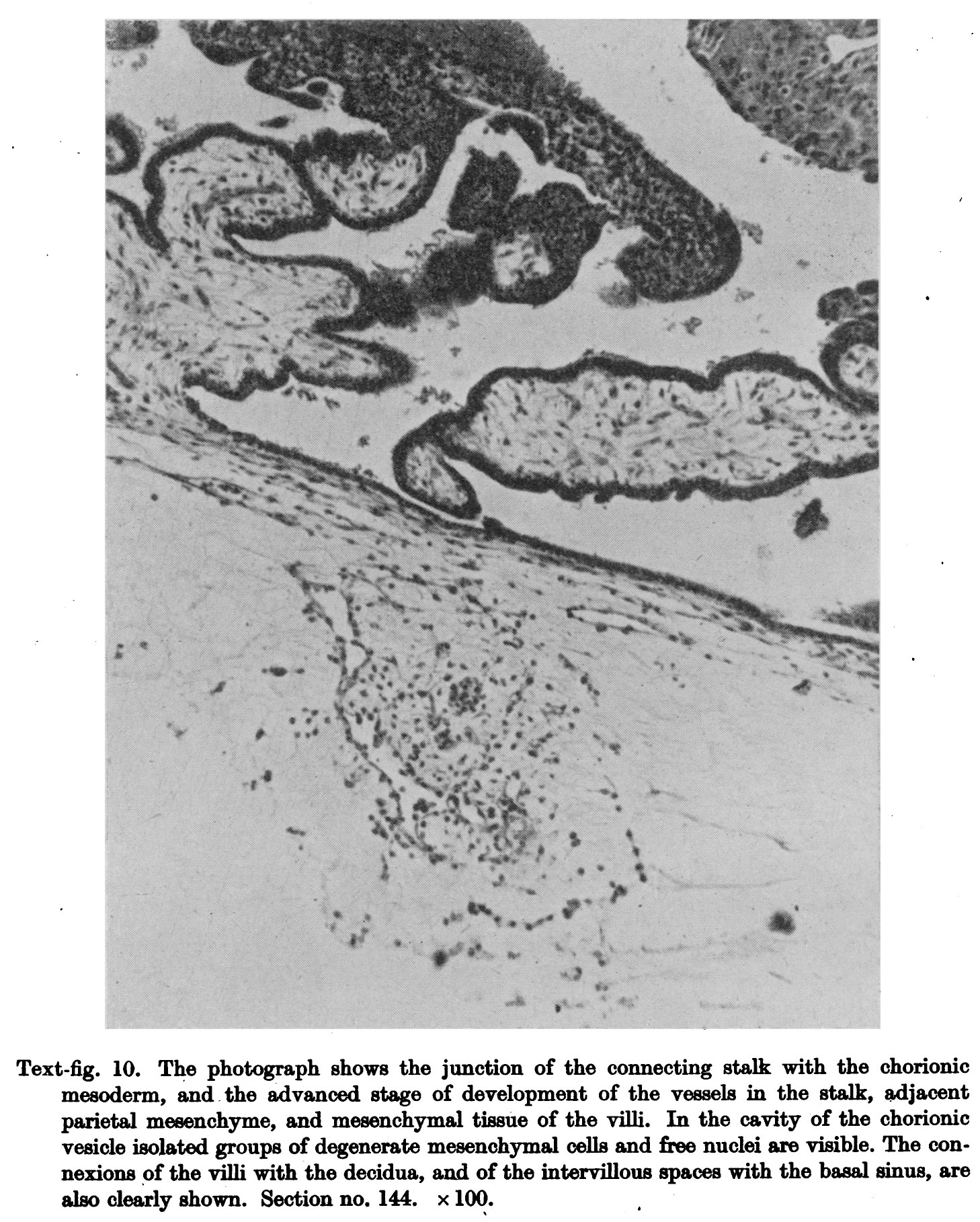 | 1,458 × 1,818 (617 KB) | Z8600021 (talk | contribs) | Text-fig. 10. The photograph shows the junction of the connecting stalk with the chorionic mesoderm, andthe advanced stage of development of the vessels in the stalk, adjacent parietal mesenchyme, and mesenchymal tissue of the In the cavity of the cho... |
You cannot overwrite this file.