File:GladstoneHamilton1941 plate04.jpg

Original file (1,280 × 1,734 pixels, file size: 325 KB, MIME type: image/jpeg)
Plate 4
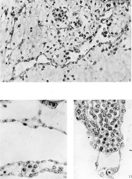
9. The section passes through the basal or chorionic end of the connecting stalk, where the supporting mesenchymal tissue resembles, and is continuous with, the parietal layer of the chorionic mesoderm. A well-developed vascular channel crosses the lower part of the photograph. The endothelial wall of this‘ channel shows various stages in the ‘rounding up’ and liberation of endothelial cell elements, some of which are seen lying free in the lumen of the vessel. Note the contrast between these cells and those in P1. 4, Hg. 11. Note also extension of solid angioblastic strands into the mesenchyme on the left. The group of epithelial cells in the upper central part of the figure probably represent a degenerate remnant of the distal portion of the allanto-enteric diverticulum. Section no. 144. x 350.
10. Section through a portion of the wall of the foregut which is shown at a lower magnification in Text-fig. 8. A small blood island is seen between the entoderm lining the floor of the foregut and the mesothelial layer covering it superficially. In the roof of the foregut opposite the blood island is a triangular space which can be traced through a series of sections. The base of the triangle is formed by entoderm; the inner and outer walls, which converge to the apex of the triangle, closely resemble the angioblastic tissue found in the chorionic mesenchyme and the basal segment of the connecting stalk. Section no. 10. x 560.
11. Segment of wall of yolk sac showing entoderm on the right side and mesothelium on the left. In the upper part of the figure the entodermal cells show signs of degeneration; in the lower part the cells appear to have been shed, leaving the endothelial wall of a Vascular space exposed. In the mesoblast is a blood island which is covered, in the greater part of its extent, by endothelium; it projects below into the vascular space and appears to be breaking through previous to discharging free erythroblasts into the lumen of the space. V x 560.
Reference
Gladstone RJ. and Hamilton WJ. A presomite human embryo (Shaw) with primitive streak and chorda canal with special reference to the development of the vascular system. (1941) Amer. J Anat. 76(1): 9-44.
Cite this page: Hill, M.A. (2024, April 27) Embryology GladstoneHamilton1941 plate04.jpg. Retrieved from https://embryology.med.unsw.edu.au/embryology/index.php/File:GladstoneHamilton1941_plate04.jpg
- © Dr Mark Hill 2024, UNSW Embryology ISBN: 978 0 7334 2609 4 - UNSW CRICOS Provider Code No. 00098G
File history
Click on a date/time to view the file as it appeared at that time.
| Date/Time | Thumbnail | Dimensions | User | Comment | |
|---|---|---|---|---|---|
| current | 16:55, 26 February 2017 |  | 1,280 × 1,734 (325 KB) | Z8600021 (talk | contribs) | |
| 16:55, 26 February 2017 |  | 1,509 × 2,044 (415 KB) | Z8600021 (talk | contribs) |
You cannot overwrite this file.