File:Gilmour1941 plate10.jpg: Difference between revisions
No edit summary |
mNo edit summary |
||
| (6 intermediate revisions by the same user not shown) | |||
| Line 1: | Line 1: | ||
==Plate X== | |||
[[:File:Gilmour1941 fig07.jpg|'''Fig. 7.''']] Embryo I, Frazer, presomite (A). Capillary-like structure in chorionic mesoderm. Heidenhain’s iron haematoxylin. x440. | |||
[[:File:Gilmour1941 fig09.jpg|'''Fig. 9.''']] 26 mm embryo. Amount of haemopoiesis in liver. Ehrlich’s {{HE}}. x155. | |||
[[:File:Gilmour1941 fig10.jpg|'''Fig. 10.''']] 444 mm foetus. Amount of haemopoiesis in liver. Ehrlich’s {{HE}}. x155. | |||
[[:File:Gilmour1941 fig11.jpg|'''Fig. 11.''']] 546 mm foetus. Amount of haemopoiesis in liver. Ehrlich’s {{HE}}. x156. | |||
{{Online Editor}} - Heidenhain's iron haematoxylin is an iron alum hematoxylin stain used for staining muscle striations and mitotic structures blue-black. Named after Rudolph Heidenhain (1834-1897) a German histologist and physiologist. (More? [[Histology Stains]]) | |||
<gallery caption="Plate 10"> | |||
File:Gilmour1941 fig07.jpg|Fig. 7 | |||
File:Gilmour1941 fig09.jpg|Fig. 9 | |||
File:Gilmour1941 fig10.jpg|Fig. 10 | |||
File:Gilmour1941 fig10.jpg|Fig. 11 | |||
</gallery> | |||
{{Gilmour1941 figures}} | |||
===Reference=== | |||
{{Ref-Gilmour1941}} | |||
{{Footer}} | |||
Latest revision as of 12:00, 17 May 2018
Plate X
Fig. 7. Embryo I, Frazer, presomite (A). Capillary-like structure in chorionic mesoderm. Heidenhain’s iron haematoxylin. x440.
Fig. 9. 26 mm embryo. Amount of haemopoiesis in liver. Ehrlich’s (Stain - Haematoxylin Eosin). x155.
Fig. 10. 444 mm foetus. Amount of haemopoiesis in liver. Ehrlich’s (Stain - Haematoxylin Eosin). x155.
Fig. 11. 546 mm foetus. Amount of haemopoiesis in liver. Ehrlich’s (Stain - Haematoxylin Eosin). x156.
Online Editor - Heidenhain's iron haematoxylin is an iron alum hematoxylin stain used for staining muscle striations and mitotic structures blue-black. Named after Rudolph Heidenhain (1834-1897) a German histologist and physiologist. (More? Histology Stains)
- Plate 10
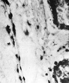
Fig. 7
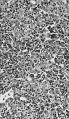
Fig. 9
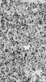
Fig. 10
Fig. 11


| Historic Disclaimer - information about historic embryology pages |
|---|
 |
Figure Links: Plate 7 | Fig. 1 | Fig. 2 | Plate 8 | Fig. 3 | Fig. 4 | Fig. 5 | Plate 9 | Fig. 6 | Fig. 8 | Plate 10 | Fig. 7 | Fig. 9 |Fig. 10 | Fig. 11 | Gilmour 1941 | Modern notes - blood | Hematopoietic and stromal cell differentiation
Reference
Gilmour JR. Normal haemopoiesis in intra-uterine and neonatal life. (1941) J. Pathol. Bacteriol. 52: 25-55.
Cite this page: Hill, M.A. (2024, May 4) Embryology Gilmour1941 plate10.jpg. Retrieved from https://embryology.med.unsw.edu.au/embryology/index.php/File:Gilmour1941_plate10.jpg
- © Dr Mark Hill 2024, UNSW Embryology ISBN: 978 0 7334 2609 4 - UNSW CRICOS Provider Code No. 00098G
File history
Click on a date/time to view the file as it appeared at that time.
| Date/Time | Thumbnail | Dimensions | User | Comment | |
|---|---|---|---|---|---|
| current | 10:29, 17 May 2018 | 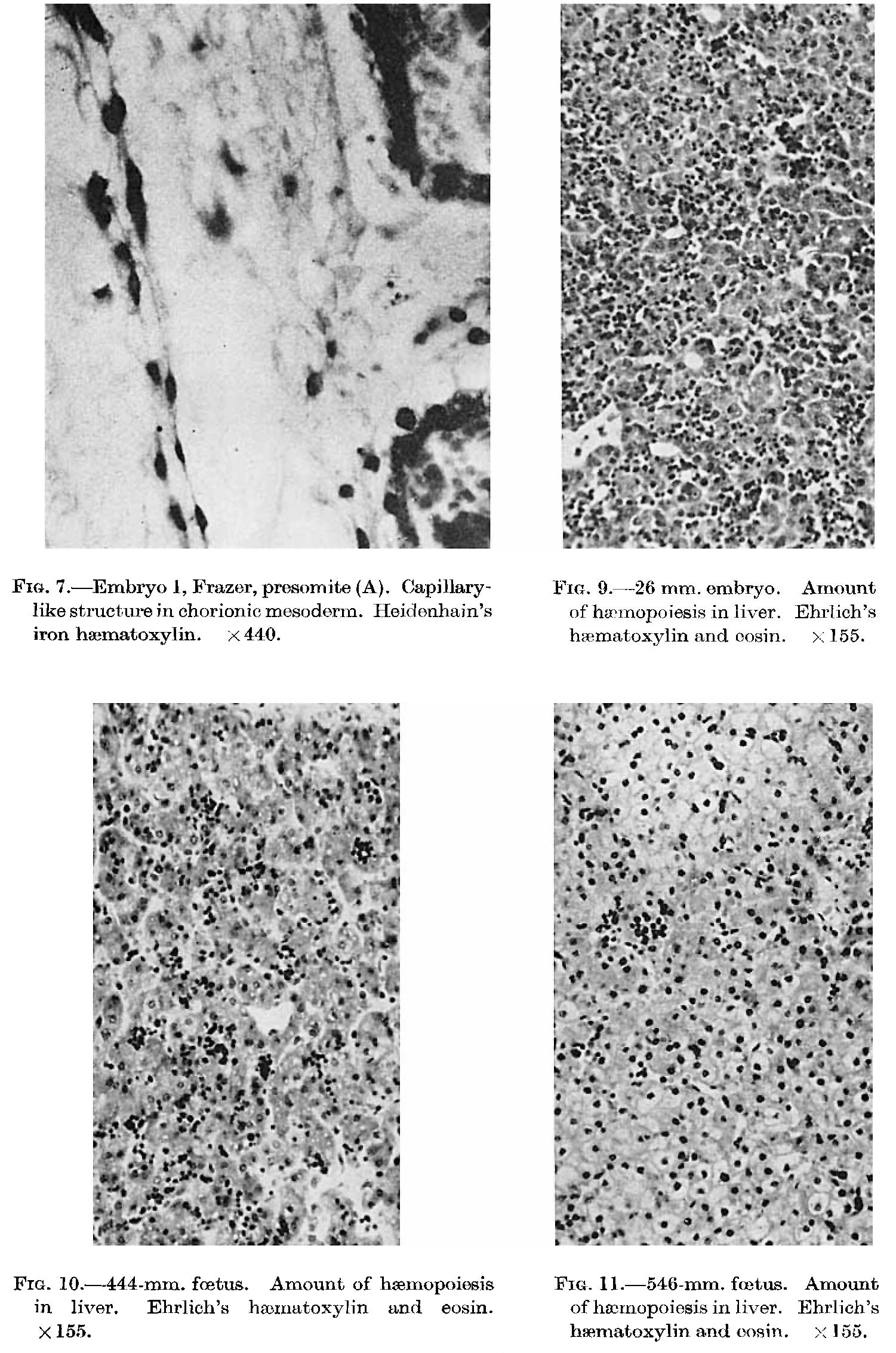 | 1,501 × 2,265 (391 KB) | Z8600021 (talk | contribs) | |
| 10:24, 17 May 2018 | 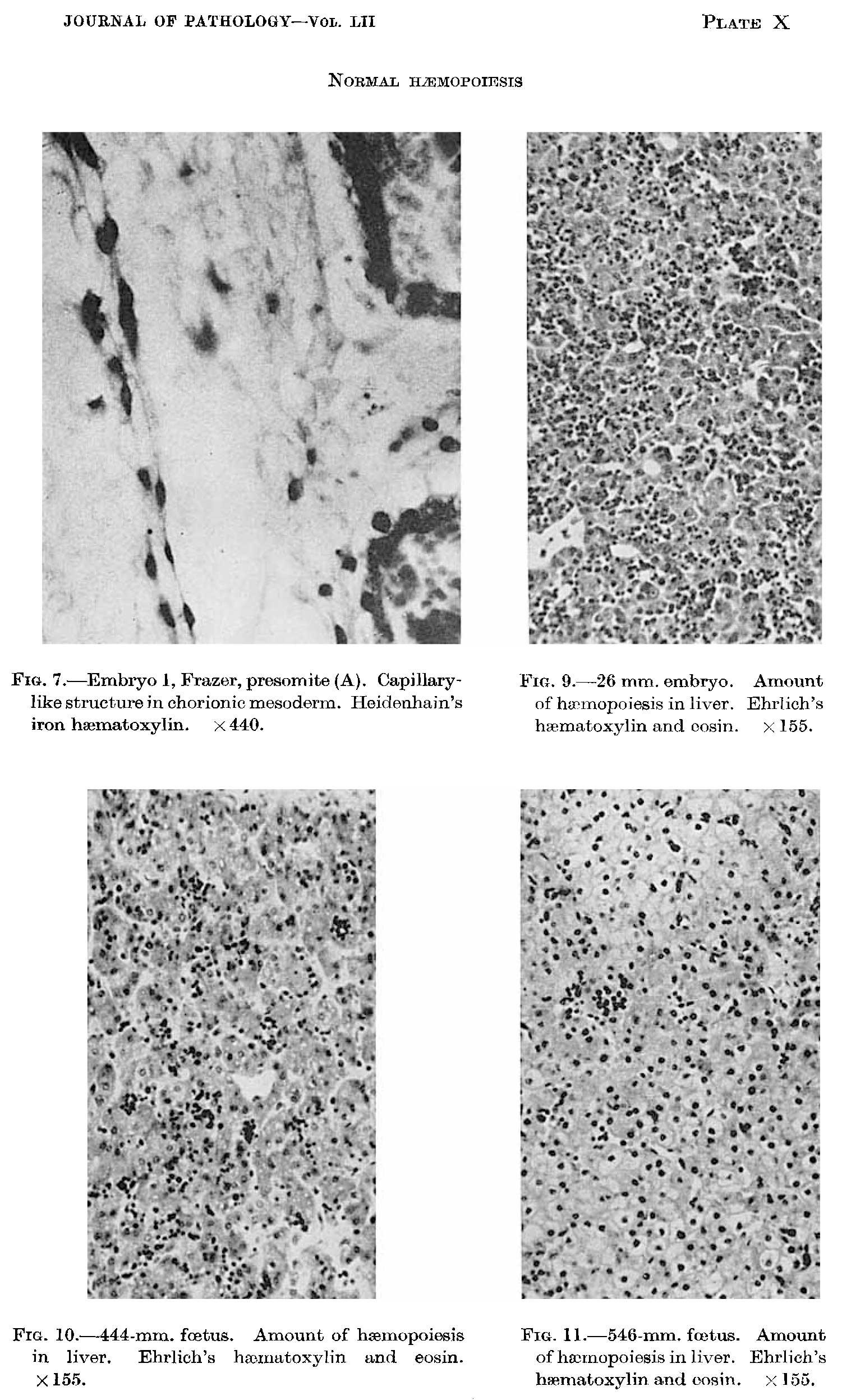 | 1,501 × 2,494 (398 KB) | Z8600021 (talk | contribs) |
You cannot overwrite this file.
File usage
The following page uses this file: