File:Gilmour1941 plate09.jpg: Difference between revisions
mNo edit summary |
mNo edit summary |
||
| (One intermediate revision by the same user not shown) | |||
| Line 1: | Line 1: | ||
==Plate IX== | ==Plate IX== | ||
'''Fig. 6.''' Embryo 4, Frazer, about 20 pairs of somites. Blood vessels in yolk sac full of primitive erythroblasts. {{HE}} Dufay process. x550. | [[:File:Gilmour1941 fig06.jpg|'''Fig. 6.''']] Embryo 4, Frazer, about 20 pairs of somites. Blood vessels in yolk sac full of primitive erythroblasts. {{HE}} Dufay process. x550. | ||
[[:File:Gilmour1941 fig08.jpg|'''Fig. 8.''']] 10 mm embryo (Frazer). Two haemocytoblasts among epithelial cells of yolk sac. Primitive erythroblasts in yolk sac vessel. Ehrlich’s {{HE}}. Dufay process. x1100. | |||
<gallery caption="Plate 9"> | |||
File:Gilmour1941 fig06.jpg|Fig. 6. | |||
File:Gilmour1941 fig08.jpg|Fig. 8. | |||
</gallery> | |||
{{Gilmour1941 figures}} | {{Gilmour1941 figures}} | ||
Latest revision as of 11:49, 17 May 2018
Plate IX
Fig. 6. Embryo 4, Frazer, about 20 pairs of somites. Blood vessels in yolk sac full of primitive erythroblasts. (Stain - Haematoxylin Eosin) Dufay process. x550.
Fig. 8. 10 mm embryo (Frazer). Two haemocytoblasts among epithelial cells of yolk sac. Primitive erythroblasts in yolk sac vessel. Ehrlich’s (Stain - Haematoxylin Eosin). Dufay process. x1100.
- Plate 9
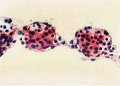
Fig. 6.
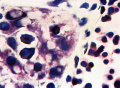
Fig. 8.


| Historic Disclaimer - information about historic embryology pages |
|---|
 |
Figure Links: Plate 7 | Fig. 1 | Fig. 2 | Plate 8 | Fig. 3 | Fig. 4 | Fig. 5 | Plate 9 | Fig. 6 | Fig. 8 | Plate 10 | Fig. 7 | Fig. 9 |Fig. 10 | Fig. 11 | Gilmour 1941 | Modern notes - blood | Hematopoietic and stromal cell differentiation
Reference
Gilmour JR. Normal haemopoiesis in intra-uterine and neonatal life. (1941) J. Pathol. Bacteriol. 52: 25-55.
Cite this page: Hill, M.A. (2024, May 7) Embryology Gilmour1941 plate09.jpg. Retrieved from https://embryology.med.unsw.edu.au/embryology/index.php/File:Gilmour1941_plate09.jpg
- © Dr Mark Hill 2024, UNSW Embryology ISBN: 978 0 7334 2609 4 - UNSW CRICOS Provider Code No. 00098G
File history
Click on a date/time to view the file as it appeared at that time.
| Date/Time | Thumbnail | Dimensions | User | Comment | |
|---|---|---|---|---|---|
| current | 10:22, 17 May 2018 | 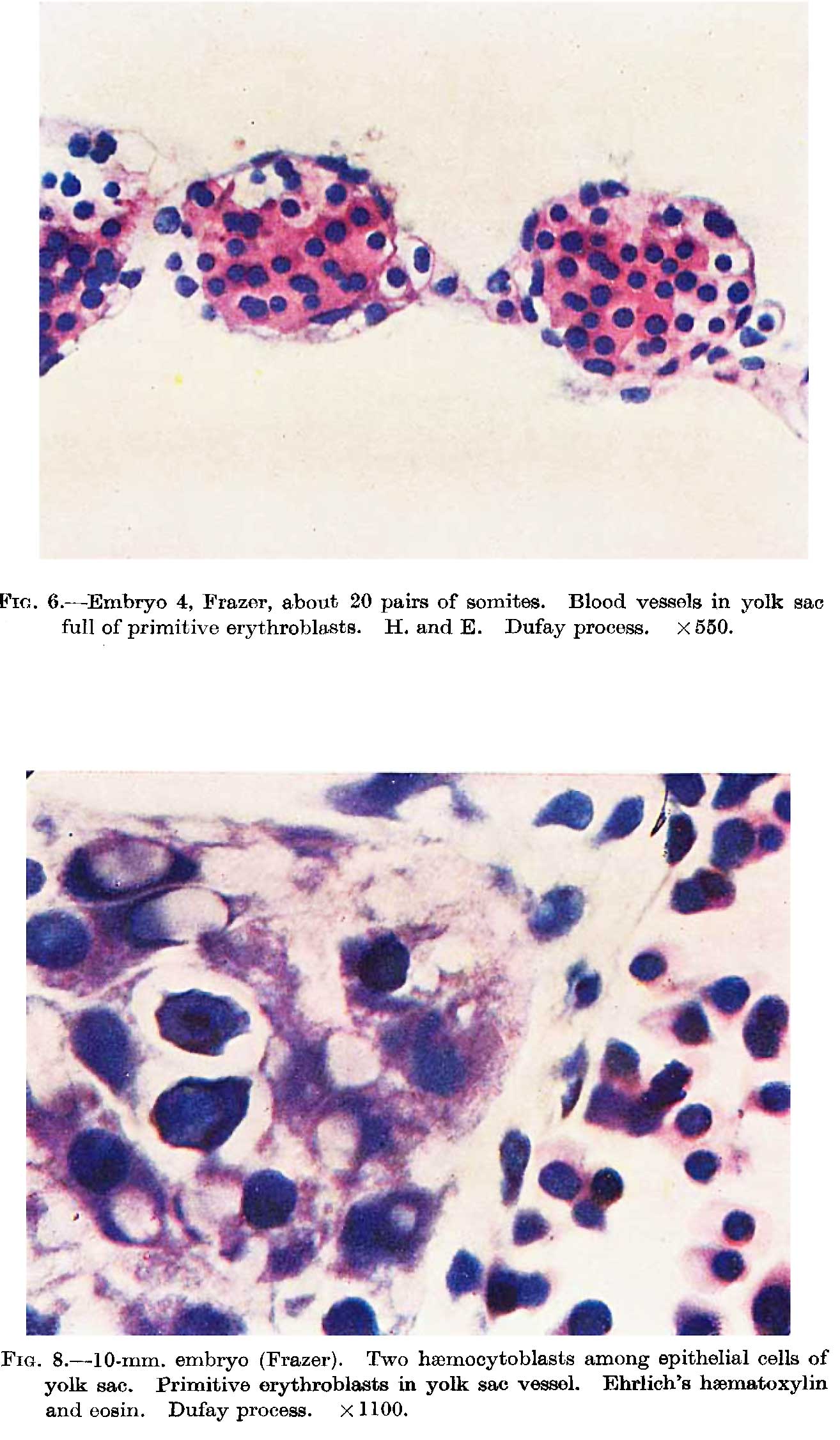 | 1,314 × 2,252 (212 KB) | Z8600021 (talk | contribs) | |
| 10:18, 17 May 2018 | 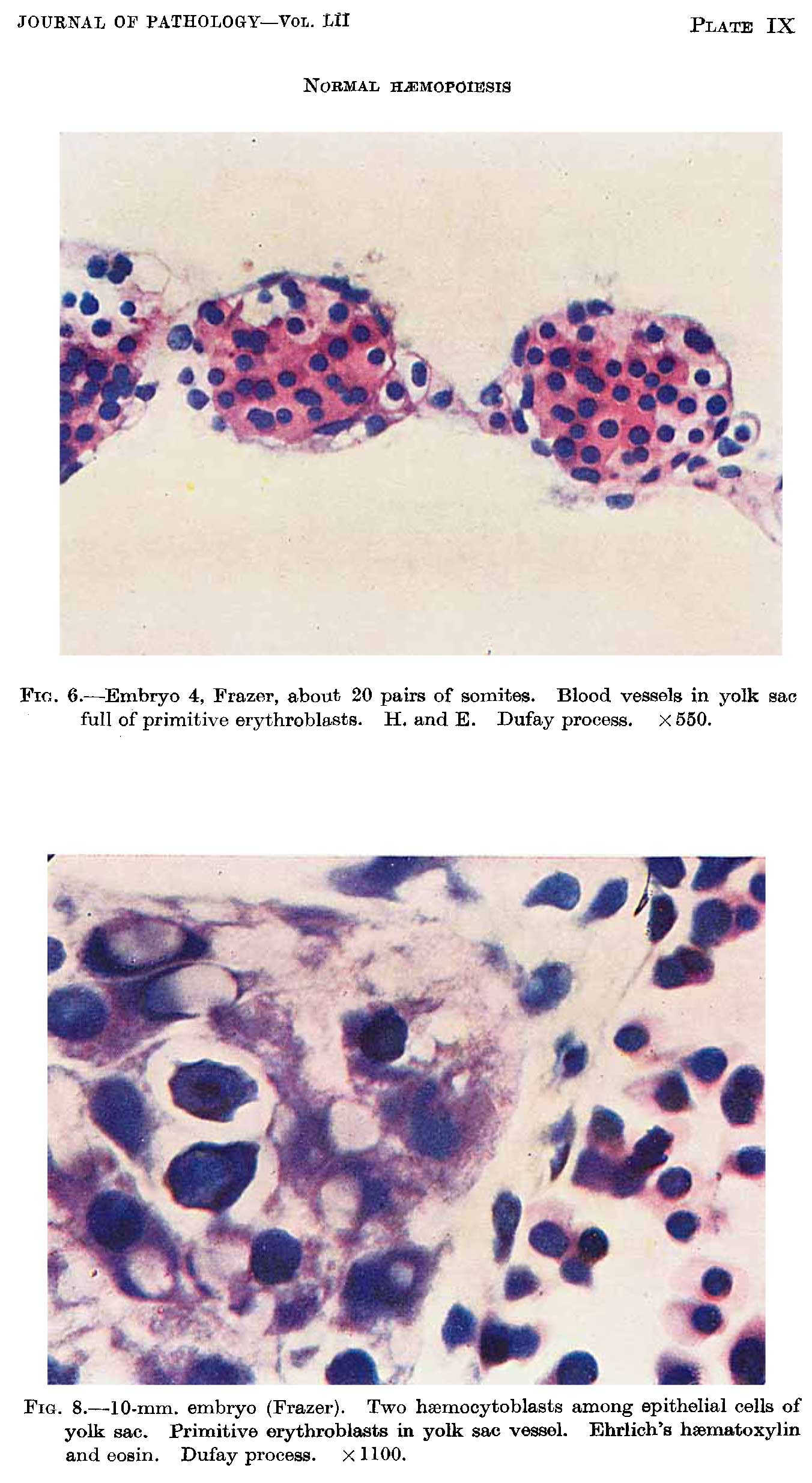 | 1,352 × 2,469 (247 KB) | Z8600021 (talk | contribs) |
You cannot overwrite this file.
File usage
The following page uses this file: