File:Frazer1926 fig07.jpg
Original file (1,229 × 996 pixels, file size: 95 KB, MIME type: image/jpeg)
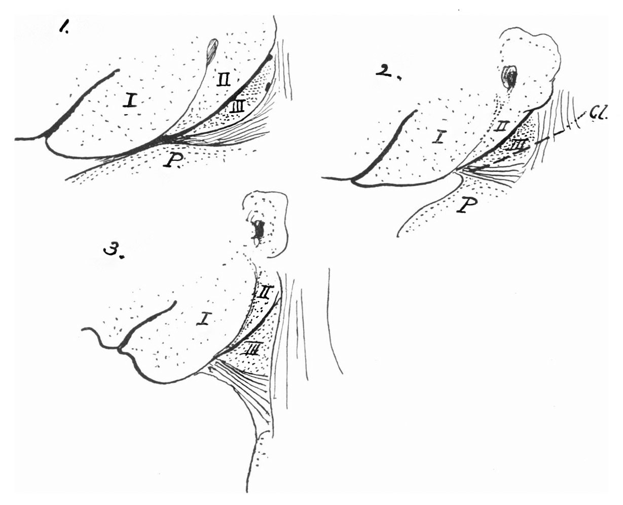
Fig. 7. Three schemes to show the situation acquired by the third arch as the neck forms
Cl. gives the general position and line of the clavicle. Other references as before. See also animation based on Fig. 7.
We have here, however, the foundation of the neck, with certain fixed points, and its formation in the second and third months runs the course that might be expected from the facts already stated. it is not necessary to add to the length of this paper by going into the details of the growth, and it will be enough for present purposes to indicate their general results in the accompanying schemes (fig. 7).
The first figure shows the general arrangement of the embryonic parts as detailed earlier in this paper, the epipericardial ridge being lined and the arches numbered. The second scheme shows the same conditions when the clavicular rudiment (Cl.) is first present and the sternomastoid is distinct. The result of the lengthening of the neck is given in the third figure. The infrahyoid muscles, fixed to the clavicle and hyoid, are drawn out on the ventral side, the sternomastoid is a fixed structure behind, and the line of flexure marks off the second and first arches in front, so that the third arch area practically corresponds with the skin covering the carotid triangle. I feel no doubt that this is quite correct in a general way, but one cannot affirm that the boundaries of the arch area and the triangle would necessarily correspond in every way. In this connection it is interesting to recall Head's "superior laryngeal" area of hyperaesthesia: it is shown in fig.8, modified from Head's original diagram, and represents a cutaneous area which might very well correspond with the skin surface of the third arch, extended under the conditions just described. It would not be justifiable, of course, to do more than suggest here such a possibility, but it may be said that Head originally proposed (Brain, Vol.xvii,1894) on theoretical grounds an association of the area with the third postoral arch.
| Historic Disclaimer - information about historic embryology pages |
|---|
 |
- Links: Fig. 1. Embryo 4-9 mm | Fig. 2. Semi-schematic pharyngeal region | Fig. 3. Embryo 8 mm | Fig.4. Embryo 10 mm | Fig. 5. Embryo 12 mm | Fig. 6. Embryo 10 mm | Fig. 7. Third Arch | Fig. 8. Laranryngeal Area of Head | Plate 1. Fig.1,3,4
Reference
Frazer JE. The disappearance of the precervical sinus. (1926) J Anat. 61(1): 132-43. PMID 17104123.
Cite this page: Hill, M.A. (2024, April 27) Embryology Frazer1926 fig07.jpg. Retrieved from https://embryology.med.unsw.edu.au/embryology/index.php/File:Frazer1926_fig07.jpg
- © Dr Mark Hill 2024, UNSW Embryology ISBN: 978 0 7334 2609 4 - UNSW CRICOS Provider Code No. 00098G
File history
Click on a date/time to view the file as it appeared at that time.
| Date/Time | Thumbnail | Dimensions | User | Comment | |
|---|---|---|---|---|---|
| current | 11:30, 28 July 2015 | | 1,229 × 996 (95 KB) | Z8600021 (talk | contribs) | |
| 11:29, 28 July 2015 |  | 1,433 × 1,102 (139 KB) | Z8600021 (talk | contribs) | ==Fig. 7. Three schemes to show the situation acquired by the third arch as the neck forms== Cl. gives the general position and line of the clavicle. Other references as before. |
You cannot overwrite this file.
File usage
The following page uses this file: