File:Frazer1926 fig05.jpg
Original file (1,000 × 643 pixels, file size: 85 KB, MIME type: image/jpeg)
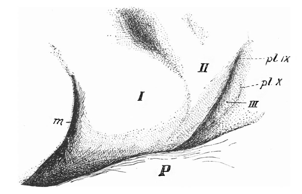
Fig. 5. Head of Embryo (12 mm)
Left side of region in an embryo of 12 mm showing the structures behind the mouth, m. I, II, III, pharyngeal arches; P, pericardium.
Ectodermal connections with placodal cysts indicated at pl. IX, pl. X.
Fig. 5 gives the surface view of the region in an embryo barely 12mm. in length. In this specimen the two placodal areas form buried cysts (cervical vesicles)related to the corresponding ganglia. Cyst X is connected with the surface still by a definite epithelial strand, enclosing a lumen at its inner end, but otherwise apparently a solid thin cord: placodal cyst IX has lost its connection with the surface, but the line formerly taken by this can, I think, be made out. The duct of this cyst disappears apparently about the 11 mm. stage. The position of these two points of final attachment to the ectoderm in the 12mm. embryo can be seen in the figure:the third arch lies, as before, between them, bounded behind by a very shallow but definite groove, and the condition is as in the 10mm. embryo with the exception that the two open ducts have been closed.
| Historic Disclaimer - information about historic embryology pages |
|---|
 |
- Links: Fig. 1. Embryo 4-9 mm | Fig. 2. Semi-schematic pharyngeal region | Fig. 3. Embryo 8 mm | Fig.4. Embryo 10 mm | Fig. 5. Embryo 12 mm | Fig. 6. Embryo 10 mm | Fig. 7. Third Arch | Fig. 8. Laranryngeal Area of Head | Plate 1. Fig.1,3,4
Reference
Frazer JE. The disappearance of the precervical sinus. (1926) J Anat. 61(1): 132-43. PMID 17104123.
Cite this page: Hill, M.A. (2024, April 27) Embryology Frazer1926 fig05.jpg. Retrieved from https://embryology.med.unsw.edu.au/embryology/index.php/File:Frazer1926_fig05.jpg
- © Dr Mark Hill 2024, UNSW Embryology ISBN: 978 0 7334 2609 4 - UNSW CRICOS Provider Code No. 00098G
File history
Click on a date/time to view the file as it appeared at that time.
| Date/Time | Thumbnail | Dimensions | User | Comment | |
|---|---|---|---|---|---|
| current | 11:19, 28 July 2015 | | 1,000 × 643 (85 KB) | Z8600021 (talk | contribs) | |
| 11:17, 28 July 2015 | 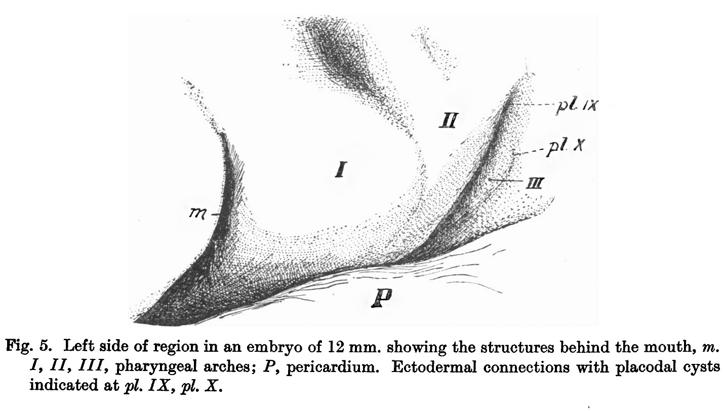 | 1,432 × 812 (141 KB) | Z8600021 (talk | contribs) | ==Fig. 5. == {{Frazer1926 figures}} |
You cannot overwrite this file.
File usage
The following page uses this file: