File:Frazer1926 fig01.jpg
Original file (1,200 × 804 pixels, file size: 137 KB, MIME type: image/jpeg)
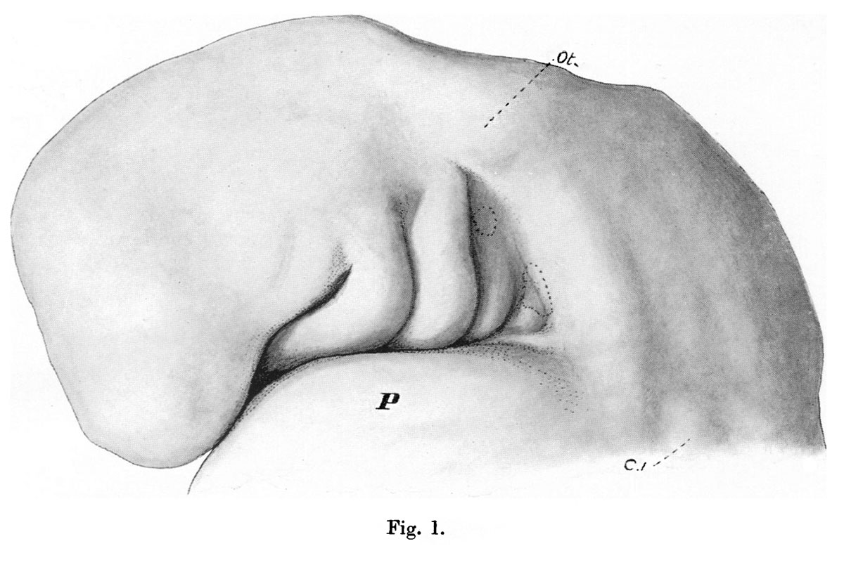
Fig. 1. Head of Embryo (4 to 9 mm)
Head of embryo of 4 to 9 mm, seen from the left. P. pericardium; C. 1, first cervical myotome; Ot.,otocyst.
The precervical sinus, a result of the disproportionate size and growth of the outer pharyngeal arches,begins to show on the surface at an early stage. Fig.1 (PI.I), the head of an embryo of less than 5 mm. in total length, gives a very good idea of this early appearance of the sinus, and of its boundaries. The third and fourth arches are visible in a triangular area, slightly depressed, in front of which the second arch is prominent and its groove very evident. Above and behind is a clearly defined broad area, especially well marked behind. The pericardium is below the sinus, but does not form its lower boundary directly: this is made by a clearly seen epipericardial ridge, which is continuous behind with the caudal boundary, and disappears from view, in front, below the lowest visible part of the third arch. The upper, hinder, and lower boundaries are thus continuous with each other round the field of the sinus, and require more particular examination.
| Historic Disclaimer - information about historic embryology pages |
|---|
 |
- Links: Fig. 1. Embryo 4-9 mm | Fig. 2. Semi-schematic pharyngeal region | Fig. 3. Embryo 8 mm | Fig.4. Embryo 10 mm | Fig. 5. Embryo 12 mm | Fig. 6. Embryo 10 mm | Fig. 7. Third Arch | Fig. 8. Laranryngeal Area of Head | Plate 1. Fig.1,3,4
Reference
Frazer JE. The disappearance of the precervical sinus. (1926) J Anat. 61(1): 132-43. PMID 17104123.
Cite this page: Hill, M.A. (2024, April 27) Embryology Frazer1926 fig01.jpg. Retrieved from https://embryology.med.unsw.edu.au/embryology/index.php/File:Frazer1926_fig01.jpg
- © Dr Mark Hill 2024, UNSW Embryology ISBN: 978 0 7334 2609 4 - UNSW CRICOS Provider Code No. 00098G
File history
Click on a date/time to view the file as it appeared at that time.
| Date/Time | Thumbnail | Dimensions | User | Comment | |
|---|---|---|---|---|---|
| current | 10:42, 28 July 2015 | | 1,200 × 804 (137 KB) | Z8600021 (talk | contribs) |
You cannot overwrite this file.
File usage
The following 2 pages use this file: