File:Frazer1922 fig06.jpg

Original file (1,000 × 728 pixels, file size: 116 KB, MIME type: image/jpeg)
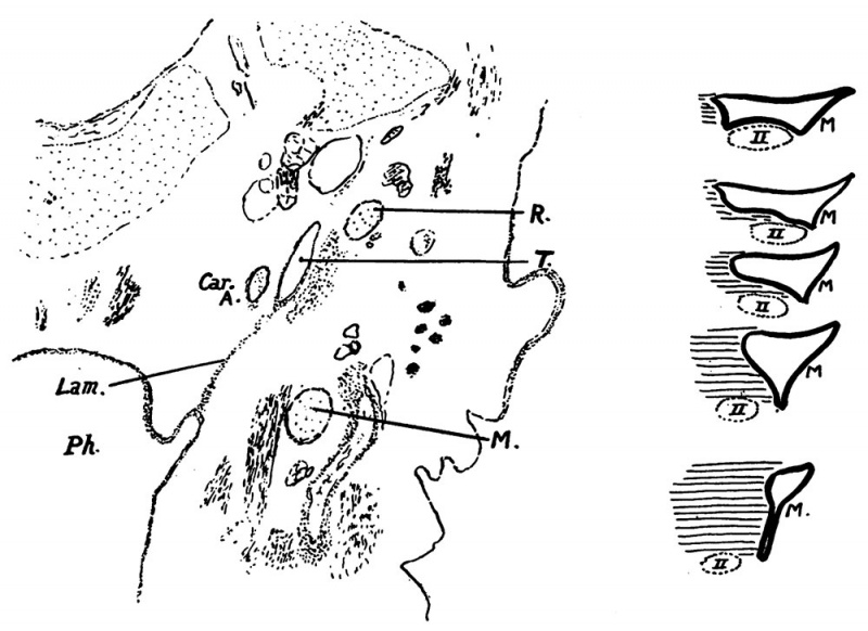
Fig 6. 35 mm Embryo
(a) Section, 35 mm. below level of patent tube. Lam. epithelial lamina connecting pharynx (Ph.) with tympanum (T); M. Meckel’s cartilage; R. Reichert’s cartilage. v
(b) Schemes to show formation of lamina; explanation in text.
Its extent is seen in the 35 mm. reconstruction in fig. 4a and its appearance on section is shown, with diagrams representing its formation, in fig. 6. The first of these diagrams is a scheme of a section across the inner part of the short recess of an early stage, parallel with the wall of the pharynx: the 3rd arch (horizontal lines) forms the hinder part of the floor and limit of the recess, the 1st arch (M) forms its front part, and the 2nd arch (dotted area) is between these two in the floor, separated from them by the 2nd and 1st grooves respectively. In the succeeding schemes the 3rd arch is seen to extend forward under the lining membrane, over the second, obliterating the 2nd groove in doing so, and gradually pushing in the hinder boundary of this part of the recess. The 2nd arch thus drops out of relation with the recess here.
| Historic Disclaimer - information about historic embryology pages |
|---|
| Pages where the terms "Historic" (textbooks, papers, people, recommendations) appear on this site, and sections within pages where this disclaimer appears, indicate that the content and scientific understanding are specific to the time of publication. This means that while some scientific descriptions are still accurate, the terminology and interpretation of the developmental mechanisms reflect the understanding at the time of original publication and those of the preceding periods, these terms, interpretations and recommendations may not reflect our current scientific understanding. (More? Embryology History | Historic Embryology Papers) |
- Links: Fig 1 | Fig 2 | Fig 3 | Fig 4 | Fig 5 | Fig 6 | 1922 Frazer | Ernest Frazer | Historic Embryology Papers | Middle Ear Development
Reference
Frazer JE. The early formations of the middle ear and eustachian tube - a criticism. (1922) J Anat. 57(1): 18-30. PMID 17103958
Cite this page: Hill, M.A. (2024, April 27) Embryology Frazer1922 fig06.jpg. Retrieved from https://embryology.med.unsw.edu.au/embryology/index.php/File:Frazer1922_fig06.jpg
- © Dr Mark Hill 2024, UNSW Embryology ISBN: 978 0 7334 2609 4 - UNSW CRICOS Provider Code No. 00098G
File history
Click on a date/time to view the file as it appeared at that time.
| Date/Time | Thumbnail | Dimensions | User | Comment | |
|---|---|---|---|---|---|
| current | 18:21, 10 January 2017 |  | 1,000 × 728 (116 KB) | Z8600021 (talk | contribs) | |
| 18:14, 10 January 2017 |  | 1,483 × 1,205 (261 KB) | Z8600021 (talk | contribs) | {{Ref-Frazer1922 figures}} |
You cannot overwrite this file.
File usage
The following 3 pages use this file:
