File:Frazer1919 fig01.jpg
From Embryology
Size of this preview: 800 × 475 pixels. Other resolution: 1,280 × 760 pixels.
Original file (1,280 × 760 pixels, file size: 123 KB, MIME type: image/jpeg)
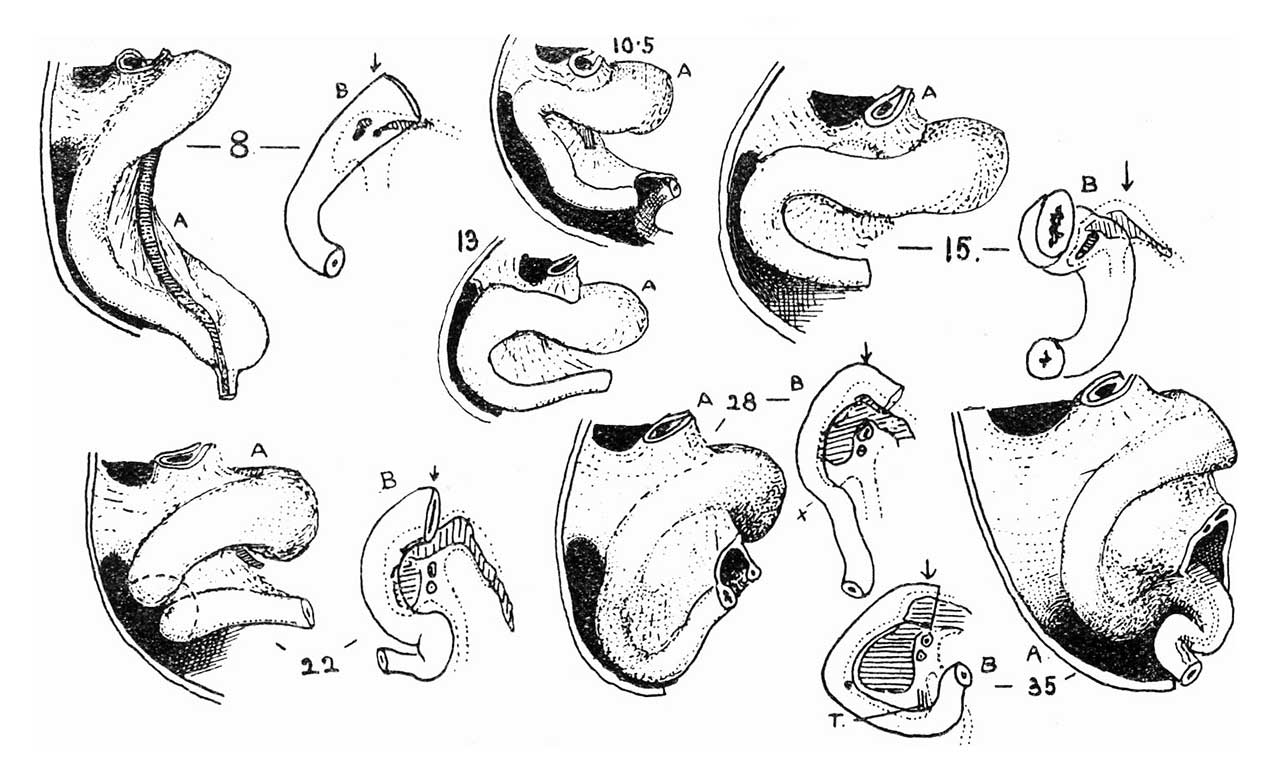
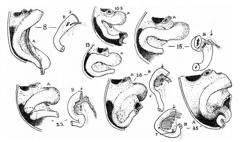
Fig. 1. The duodenum throughout the “first stage” of intestinal development
A, side view, B, front view. The numerals give the size of embryo in millimetres. See text.
Reference
Frazer JE. The formation of the duodenal curve. J Anat. 1919 53(4):292-7. PMID 17103870
Cite this page: Hill, M.A. (2024, April 26) Embryology Frazer1919 fig01.jpg. Retrieved from https://embryology.med.unsw.edu.au/embryology/index.php/File:Frazer1919_fig01.jpg
- © Dr Mark Hill 2024, UNSW Embryology ISBN: 978 0 7334 2609 4 - UNSW CRICOS Provider Code No. 00098G
File history
Click on a date/time to view the file as it appeared at that time.
| Date/Time | Thumbnail | Dimensions | User | Comment | |
|---|---|---|---|---|---|
| current | 13:07, 15 June 2018 | | 1,280 × 760 (123 KB) | Z8600021 (talk | contribs) | |
| 13:06, 15 June 2018 | 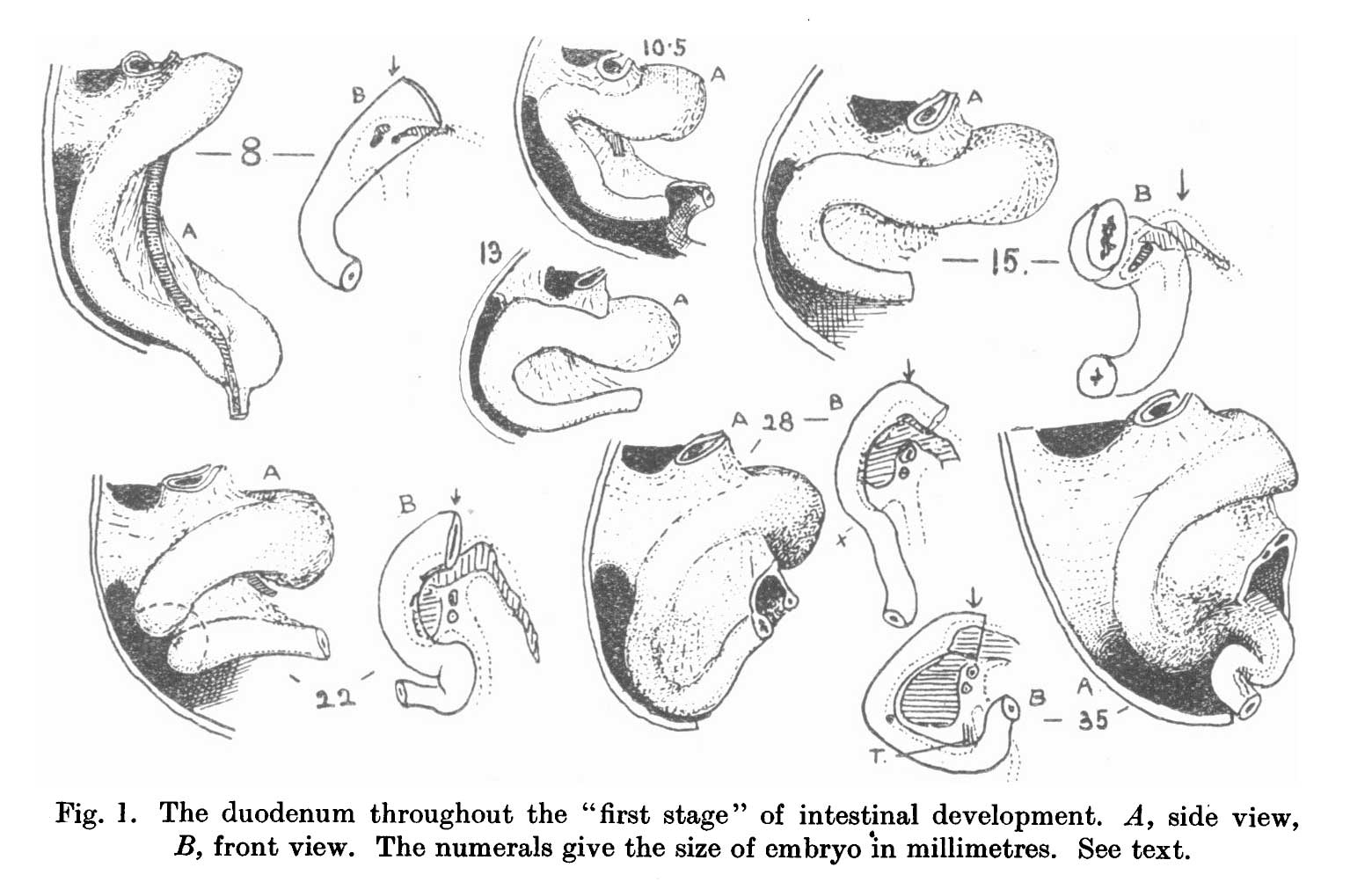 | 1,560 × 1,003 (137 KB) | Z8600021 (talk | contribs) | ===Reference=== {{Ref-Frazer1919}} {{Footer}} |
You cannot overwrite this file.
File usage
The following page uses this file: