File:Frazer1910 fig07d.jpg: Difference between revisions
({{Frazer1910 figures}}) |
mNo edit summary |
||
| Line 1: | Line 1: | ||
==Fig. 7D. Reconstruction model embryo 22 mm== | |||
D gives the outline of the hyoid, thyroid, and inter-thyroid lamina, and cricoid from the front. | |||
The numerous remaining structures in the last three models, with the other points that are evident in the earlier ones, will be described as they come into the account of the growth of the larynx. | |||
A short general consideration of the floor of the pharynx can conveniently precede the description of the laryngeal development, with the object of showing that five arches are represented in this floor. | |||
<gallery> | |||
Frazer1910 fig07a.jpg|7 A side view | |||
Frazer1910 fig07b.jpg|7 B side view of the deeper structures | |||
Frazer1910 fig07c.jpg|7 C thyroid ala | |||
Frazer1910 fig07d.jpg|7 D from the front. | |||
</gallery> | |||
{{Frazer1910 figures}} | {{Frazer1910 figures}} | ||
Latest revision as of 09:27, 11 January 2017
Fig. 7D. Reconstruction model embryo 22 mm
D gives the outline of the hyoid, thyroid, and inter-thyroid lamina, and cricoid from the front.
The numerous remaining structures in the last three models, with the other points that are evident in the earlier ones, will be described as they come into the account of the growth of the larynx.
A short general consideration of the floor of the pharynx can conveniently precede the description of the laryngeal development, with the object of showing that five arches are represented in this floor.
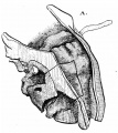
7 A side view
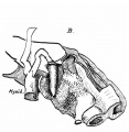
7 B side view of the deeper structures
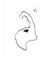
7 C thyroid ala
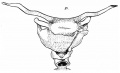
7 D from the front.


| Historic Disclaimer - information about historic embryology pages |
|---|
 |
- Links: fig 1 | fig 2 | fig 3 | fig 4 | fig 5 | fig 6 | fig 7 | fig 8 | fig 9 | fig 10 | fig 11 | fig 12 | fig 13 | fig 14 | fig 15 | fig 16 | fig 17 | fig 18 | fig 19 | 1910 Frazer | Historic Embryology Papers | Respiratory System Development
Reference
Frazer JE. Development of the larynx. (1910) J Anat. 44: 156-191. PMID 17232839
Cite this page: Hill, M.A. (2024, May 21) Embryology Frazer1910 fig07d.jpg. Retrieved from https://embryology.med.unsw.edu.au/embryology/index.php/File:Frazer1910_fig07d.jpg
- © Dr Mark Hill 2024, UNSW Embryology ISBN: 978 0 7334 2609 4 - UNSW CRICOS Provider Code No. 00098G
File history
Click on a date/time to view the file as it appeared at that time.
| Date/Time | Thumbnail | Dimensions | User | Comment | |
|---|---|---|---|---|---|
| current | 09:24, 11 January 2017 |  | 1,036 × 634 (72 KB) | Z8600021 (talk | contribs) | {{Frazer1910 figures}} |
You cannot overwrite this file.
File usage
The following 6 pages use this file: