File:Foster114.jpg
From Embryology

Size of this preview: 406 × 600 pixels. Other resolution: 725 × 1,071 pixels.
Original file (725 × 1,071 pixels, file size: 135 KB, MIME type: image/jpeg)
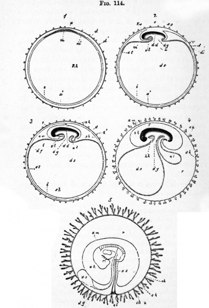
Fig. 114. Five diagrammatic figures illustrating the formation of the foetal membranes of a mammal
(From Kolliker.)
- In 1, 2, 3, 4 the embryo is represented in longitudinal section.
- Ovurn with zona pellucicla, blastodermic vesicle, and embryonic area.
- Ovum with commencing formation of umbilical vesicle and amnion.
- Ovum with amnion about to close, and commencing allantois.
- Ovum with villous subzonal membrane, larger allantois, and mouth and anus.
- Ovum in which the mesoblast of the allantois has extended round the inner surface of the subzonal membrane and united with it to form the chorion. The cavity of the allantois is aborted. This fig. is a diagram of an early human ovum.
Legend
- d. zona radiata ; d and sz. processes of zona ; sh. subzonal membrane, outer fold of amnion, false amnion ; ch. chorion ; ch. z. chorionic villi ; am. amnion ; ks. head-fold of amnion ; ss. tailfold of amnion ; a. epiblast of embryo ; a. epiblast of non-embryonic part of the blastodermic vesicle ; m. embryonic mesoblast ; m'. non-embryonic mesoblast ; df. area vasculosa ; st. sinus terminalis; dd. embryonic hypoblast; i. non-embryonic hypoblast ; kh. cavity of blastodermic vesicle, the greater part of which becomes the cavity of umbilical vesicle ds. ; dg. stalk of umbilical vesicle ; al. allantois ; e. embryo ; r. space between chorion and amnion containing albuminous fluid ; vl. ventral body wall ; hh. pericardial cavity.
Reference
Foster, M., Balfour, F. M., Sedgwick, A., & Heape, W. (1883). The Elements of Embryology. (2nd ed.). London: Macmillan and Co.
- Volume 1 - The History of the Chick: Egg structure and incubation beginning | Summary whole incubation | First day | Second day - first half | Second day - second half | Third day | Fourth day | Fifth day | Sixth day to incubation end | Figures 1
- Volume 2 - The History of the Mammalian Embryo: General Development | Embryonic Membranes and Yolk-Sac | Organs from Epiblast | Organs from Mesoblast | Alimentary Canal | Appendix | Figures 2
| Historic Disclaimer - information about historic embryology pages |
|---|
| Pages where the terms "Historic" (textbooks, papers, people, recommendations) appear on this site, and sections within pages where this disclaimer appears, indicate that the content and scientific understanding are specific to the time of publication. This means that while some scientific descriptions are still accurate, the terminology and interpretation of the developmental mechanisms reflect the understanding at the time of original publication and those of the preceding periods, these terms, interpretations and recommendations may not reflect our current scientific understanding. (More? Embryology History | Historic Embryology Papers) |
File history
Click on a date/time to view the file as it appeared at that time.
| Date/Time | Thumbnail | Dimensions | User | Comment | |
|---|---|---|---|---|---|
| current | 18:10, 12 January 2011 |  | 725 × 1,071 (135 KB) | S8600021 (talk | contribs) | FIG. 114. FIVE DIAGRAMMATIC FIGURES ILLUSTRATING THE FORMATION OF THE FOETAL MEMBRANES OF A MAMMAL. (From Kolliker.) In 1, 2, 3, 4 the embryo is represented in longitudinal section. 1. Ovurn with zona pellucicla, blastodermic vesicle, and embryonic area |
You cannot overwrite this file.
File usage
The following 2 pages use this file:
