File:Foster035.jpg
Foster035.jpg (246 × 253 pixels, file size: 13 KB, MIME type: image/jpeg)
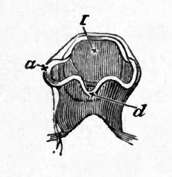
FIG. 35. HEAD OF A CHICK AT THE END OF THE SECOND DAY VIEWED FROM BELOW AS A TRANSPARENT OBJECT.
(Copied from Huxley).
I. first cerebral vesicle, a. optic vesicle, d. infundibulum.
The specimen shews the formation of the optic vesicles (a), as outgrowths from the 1st cerebral vesicle or vesicle of the 3rd ventricle, so that the optic vesicles and vesicle of the 3rd ventricle at first freely communicated with each other, and also the growth of the lower wall of the vesicle of the 3rd ventricle into a process which becomes the infundibulum (d).
| Historic Disclaimer - information about historic embryology pages |
|---|
 |
Reference
Foster, M., Balfour, F. M., Sedgwick, A., & Heape, W. (1883). The Elements of Embryology. (2nd ed.). London: Macmillan and Co.
The Elements of Embryology (1883)
File history
Click on a date/time to view the file as it appeared at that time.
| Date/Time | Thumbnail | Dimensions | User | Comment | |
|---|---|---|---|---|---|
| current | 08:51, 11 January 2011 | | 246 × 253 (13 KB) | S8600021 (talk | contribs) | FIG. 35. HEAD OF A CHICK AT THE END OF THE SECOND DAY VIEWED FROM BELOW AS A TRANSPARENT OBJECT. (Copied from Huxley). I. first cerebral vesicle, a. optic vesicle, d. infundibulum. The specimen shews the formation of the optic vesicles (a), as outgr |
You cannot overwrite this file.
File usage
The following 2 pages use this file: