File:Foster010.jpg
Original file (971 × 497 pixels, file size: 57 KB, MIME type: image/jpeg)
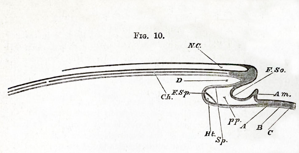
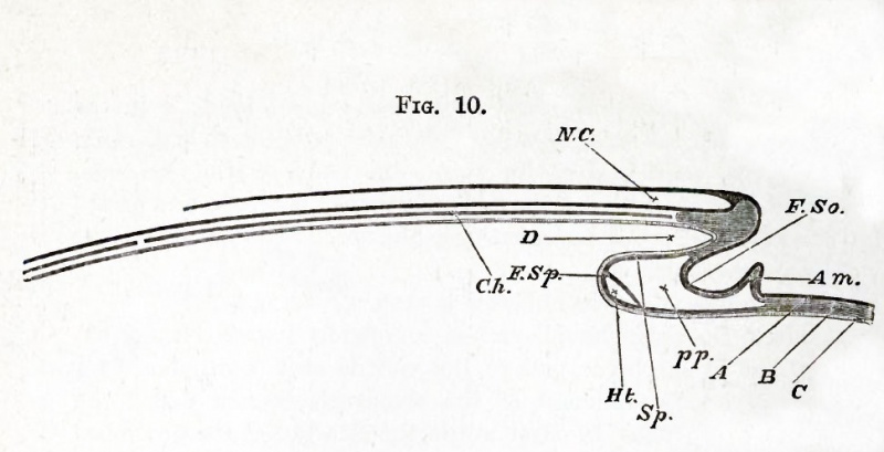
FIG. 10. DIAGRAMMATIC LONGITUDINAL SECTION THROUGH THE AXIS OF AN EMBRYO.
The section is supposed to be made at a time when the headfold has commenced but the tail-fold has not yet appeared. F. So. fold of the somatopleure. F. Sp. fold of the splanchnopleure.
The line of reference F. So. is placed in the lower bay, outside the embryo. The line of D is placed in the upper bay inside the embryo ; this will remain as the alimentary canal. Both folds (F. So., F. Sp.) are parts of the head- fold, and are to be thought of as continually travelling onwards (to the left) as development proceeds. pp. space between somatopleure and splanchnopleure : pleuro peritoneal cavity. Am. commencing (head) fold of the amnion.
A fuller explanation is given under Fig. 29.
| Historic Disclaimer - information about historic embryology pages |
|---|
 |
Reference
Foster, M., Balfour, F. M., Sedgwick, A., & Heape, W. (1883). The Elements of Embryology. (2nd ed.). London: Macmillan and Co.
The Elements of Embryology (1883)
File history
Click on a date/time to view the file as it appeared at that time.
| Date/Time | Thumbnail | Dimensions | User | Comment | |
|---|---|---|---|---|---|
| current | 16:00, 8 January 2011 | | 971 × 497 (57 KB) | S8600021 (talk | contribs) | {{Template:Foster 1883 Figures}} |
You cannot overwrite this file.
File usage
The following 2 pages use this file: