File:Foster009ln.jpg

Original file (977 × 1,038 pixels, file size: 113 KB, MIME type: image/jpeg)
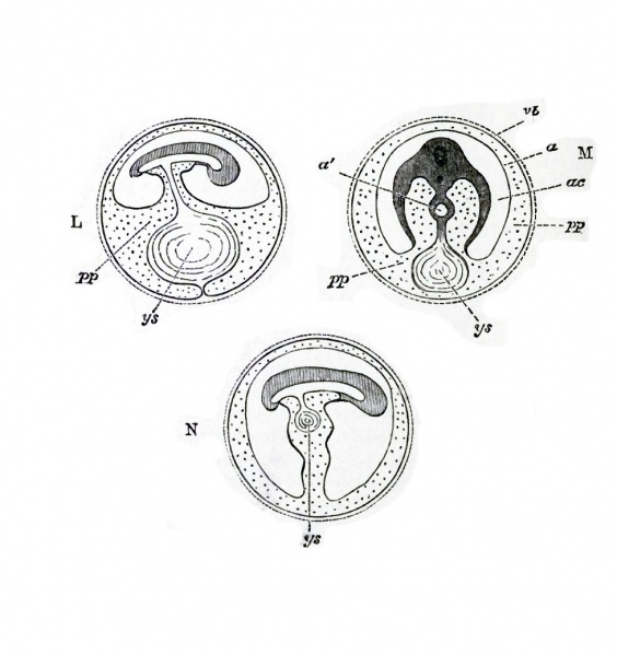
Fig. 9, A to N forms a series of purely diagrammatic representations introduced to facilitate the comprehension of the manner in which the body of the embryo is formed, and of the various relations of the yolk-sac, amnion and allantois.
In L the splanchnopleure has completely invested the yolksac, but at the lower pole of the yolk is still continuous with that peripheral remnant of the somatopleure now called the serous membrane. In other words, the cleavage of the mesoblast has been carried all round the yolk (ys) except just at the lower pole.
In M the cleavage has been carried through the pole itself ; the peripheral portion of the splanchnopleure forms a complete investment of the yolk, quite unconnected with the peripheral portion of the somatopleure, which now exists as a continuous membrane lining the interior of the shell. The yolk-sac (ys) is therefore quite loose in the pleuroperitoneal cavity, being connected only with the alimentary canal (a') by a solid pedicle.
Lastly, in N the yolk-sac (ys} is shewn being withdrawn into the cavity of the body of the embryo. The allantois is as before, for the sake of simplicity, omitted ; its pedicle would of course lie by the side of ys in the somatic stalk marked by the usual dotted shading.
| Historic Disclaimer - information about historic embryology pages |
|---|
 |
Reference
Foster, M., Balfour, F. M., Sedgwick, A., & Heape, W. (1883). The Elements of Embryology. (2nd ed.). London: Macmillan and Co.
The Elements of Embryology (1883)
File history
Click on a date/time to view the file as it appeared at that time.
| Date/Time | Thumbnail | Dimensions | User | Comment | |
|---|---|---|---|---|---|
| current | 15:49, 8 January 2011 | | 977 × 1,038 (113 KB) | S8600021 (talk | contribs) |
You cannot overwrite this file.
File usage
The following 2 pages use this file: