File:Florian1930-text-fig02.jpg
Florian1930-text-fig02.jpg (394 × 377 pixels, file size: 20 KB, MIME type: image/jpeg)
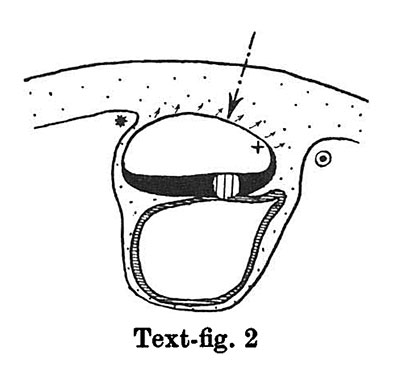
Text-fig 2. Schemes of the chief stages in the development of the human embryo
In stage 2 (text-fig. 2) the mesoderm covering the whole yolk-sac and the cranial end of the amniotic cavity has taken on a different structure, and is becoming transformed into a "mesothelium." The cells of the mesoderm in this region are connected with the cells of the amniotic ectoderm and with those of the yolk-sac entoderm by means of cytodesmata, and some of the cells of the yolk-sac entoderm appear to have entered the mesoderm. The transition of the mesodermal layer which covers the cranial end of the amnion into the chorionic mesoderm (text-fig. 2*) represents the cranial limit of the connecting stalk, which first begins to be formed in this stage. The first traces of the stalk appear, therefore, in the stage when the primitive streak primordium is represented by a localised fusion of the embryonal ectoderm with the entoderm of the yolk-sac (embryo OP v.Mollendorff).The yolk-sac (in this stage) runs out towards the connecting stalk in the form of a slight diverticulum. Already in this stage v. Mollendorff described (1921,1925) an active extension of the amniotic cavity towards the stalk tissue (in a dorsal and caudal direction from the embryonal plate). If we regard the caudal limit of the connecting stalk (in text-figs. 1-8) as a fixed point, we may say that the embryonal plate grows in the cranial direction, while the amniotic cavity expands caudally and dorsally. The yolk-sac correspondingly grows forwards below the embryonic plate almost up to the cranial end of the latter, and also grows in the caudal direction. Its growth in this direction, however, encounters the resistance of the connecting stalk tissue and so, not being able to extend freely, it grows towards the connecting stalk in the form of a diverticulum.
Already in this stage we can distinguish two parts in the connecting stalk which I propose to term the amnio-embryonal stalk and the umbilical stalk respectively. The amnio-embryonal stalk (between + and ʘ in text-figs. 1-8) attaches the embryonal formation (amnio-embryonal and yolk-sac vesicles) as a whole to the chorionic mesoderm and continues on the one hand into the amniotic mesoderm, and on the other into the umbilical stalk.
The umbilical stalk (between + and - in text-figs. 2-8) represents a partial continuation of the amnio-embryonal stalk; on its cranial surface it is covered by the stalk ectoderm which is continuous with the ectoderm of the embryonic plate and with that of the amnion, and its whole surface can only be seen if the amnion is opened up. The most important difference between the amnio-embryonal stalk and the umbilical stalk lies in the fact that the latter is in all stages covered on its cranial surface by ectoderm and that it is transformed into the umbilical cord during later stages of development. The amnio-embryonal stalk on the other hand persists for a time but finally disappears when the amniotic mesoderm fuses with that of the chorion. The axis of the connecting stalk (marked by an arrow) begins already in stage 2 (text-fig.2) to form an acute angle (openc audally) with the embryonic plate and has moved slightly towards the caudal end of the embryo.
Ectoderm black; entoderm lined horizontally, primitive streak vertically, head process and chorda plate obliquely. Mesoderm dotted. The axis of the connecting stalk is marked by an arrow. The limits of the connecting stalk are marked by * and ʘ, the limits of the umbilical stalk by + and ʘ). The direction of the extension of the amniotic cavity towards the connecting stalk is marked by arrows. The parts of the allantois and of the axis of the connecting stalk situated out of the median plane are finely dotted. A. Connecting stalk; B. umbilical stalk; Cl.m. cloacal membrane.
| Historic Disclaimer - information about historic embryology pages |
|---|
 |
- Links: Text-fig 1 | Text-fig 2 | Text-fig 3 | Text-fig 4 | Text-fig 5 | Text-fig 6 | Text-fig 7 | Text-fig 1-7 | Text-fig 8 | Text-fig 9 | Text-fig 8-9 | Florian 1930 | Historic Embryology Papers
Reference
Florian J. The formation of the connecting stalk and the extension of the amniotic cavity towards the tissue of the connecting stalk in young human embryos. (1930) J. Anat., 64: 454-476.
Cite this page: Hill, M.A. (2024, April 27) Embryology Florian1930-text-fig02.jpg. Retrieved from https://embryology.med.unsw.edu.au/embryology/index.php/File:Florian1930-text-fig02.jpg
- © Dr Mark Hill 2024, UNSW Embryology ISBN: 978 0 7334 2609 4 - UNSW CRICOS Provider Code No. 00098G
File history
Click on a date/time to view the file as it appeared at that time.
| Date/Time | Thumbnail | Dimensions | User | Comment | |
|---|---|---|---|---|---|
| current | 18:58, 23 September 2015 | | 394 × 377 (20 KB) | Z8600021 (talk | contribs) |
You cannot overwrite this file.
File usage
The following 3 pages use this file: