File:Flecker1932 fig08.jpg
Flecker1932_fig08.jpg (300 × 363 pixels, file size: 18 KB, MIME type: image/jpeg)
Fig. 6. Age 21. Sesamoids at heads of first, second, third and fifth metacarpals. Large sesamoid over centre of third metacarpal.
Fig. 7. Male, age 18. Epiphyses along perineal margin at apex of perineal triangle.
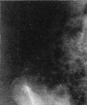
Fig. 8. Female, age 14. Epiphysis for anterior superior iliac spine.
Fig. 9. Female, age 17. Epiphysis for anterior superior iliac spine.
Fig. 10. Female, aged 2 years and 3 months. Two centres present for first cuneiform.
Cite this page: Hill, M.A. (2024, May 20) Embryology Flecker1932 fig08.jpg. Retrieved from https://embryology.med.unsw.edu.au/embryology/index.php/File:Flecker1932_fig08.jpg
- © Dr Mark Hill 2024, UNSW Embryology ISBN: 978 0 7334 2609 4 - UNSW CRICOS Provider Code No. 00098G
File history
Click on a date/time to view the file as it appeared at that time.
| Date/Time | Thumbnail | Dimensions | User | Comment | |
|---|---|---|---|---|---|
| current | 10:49, 9 February 2020 | | 300 × 363 (18 KB) | Z8600021 (talk | contribs) |
You cannot overwrite this file.
File usage
The following 7 pages use this file: