File:Flecker1932 fig06.jpg: Difference between revisions
No edit summary |
mNo edit summary |
||
| (One intermediate revision by the same user not shown) | |||
| Line 1: | Line 1: | ||
==Fig. 6. Age 21. Sesamoids at heads of first, second, third and fifth metacarpals== | |||
Large sesamoid over centre of third metacarpal. | |||
<gallery> | |||
File:Flecker1932 fig06.jpg|Fig. 6. Age 21. Sesamoids at heads of first, second, third and fifth metacarpals. | |||
File:Flecker1932 fig07.jpg|Fig. 7. Male, age 18. Epiphyses along perineal margin at apex of perineal triangle. | |||
File:Flecker1932 fig08.jpg|Fig. 8. Female, age 14. Epiphysis for anterior superior iliac spine. | |||
File:Flecker1932 fig09.jpg|Fig. 9. Female, age 17. Epiphysis for anterior superior iliac spine. | |||
File:Flecker1932 fig10.jpg|Fig. 10. Female, aged 2 years and 3 months. Two centres present for first cuneiform. | |||
</gallery> | |||
===Reference=== | |||
{{Ref-Flecker1932}} | |||
{{Footer}} | |||
[[Category:Historic Embryology]][[Category:1930's]][[Category:Bone]][[Category:X-ray]] | |||
[[Category:Draft]] | |||
Latest revision as of 10:54, 9 February 2020
Fig. 6. Age 21. Sesamoids at heads of first, second, third and fifth metacarpals
Large sesamoid over centre of third metacarpal.
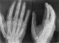
Fig. 6. Age 21. Sesamoids at heads of first, second, third and fifth metacarpals.
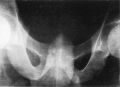
Fig. 7. Male, age 18. Epiphyses along perineal margin at apex of perineal triangle.
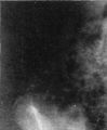
Fig. 8. Female, age 14. Epiphysis for anterior superior iliac spine.
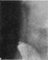
Fig. 9. Female, age 17. Epiphysis for anterior superior iliac spine.
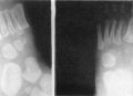
Fig. 10. Female, aged 2 years and 3 months. Two centres present for first cuneiform.


Reference
Flecker H. (1932). Roentgenographic observations of the times of appearance of epiphyses and their fusion with the diaphyses. (1932) J Anat. 67: 118-164.3 PMID 17104405
Cite this page: Hill, M.A. (2024, May 18) Embryology Flecker1932 fig06.jpg. Retrieved from https://embryology.med.unsw.edu.au/embryology/index.php/File:Flecker1932_fig06.jpg
- © Dr Mark Hill 2024, UNSW Embryology ISBN: 978 0 7334 2609 4 - UNSW CRICOS Provider Code No. 00098G
File history
Click on a date/time to view the file as it appeared at that time.
| Date/Time | Thumbnail | Dimensions | User | Comment | |
|---|---|---|---|---|---|
| current | 10:49, 9 February 2020 |  | 700 × 513 (106 KB) | Z8600021 (talk | contribs) |
You cannot overwrite this file.
File usage
The following 7 pages use this file: