File:Fetal ultrasound ductal arch 01.jpg
Fetal_ultrasound_ductal_arch_01.jpg (800 × 533 pixels, file size: 27 KB, MIME type: image/jpeg)
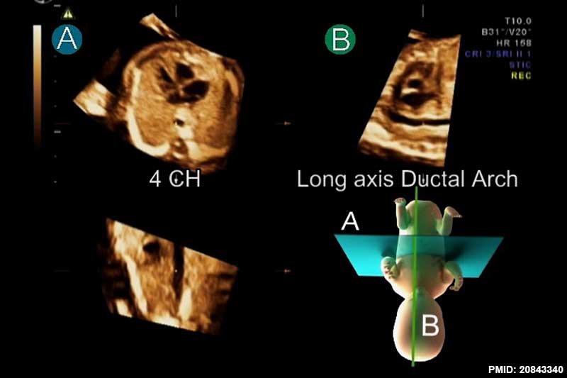
Fetal Ultrasound of the Ductal Arch
The ductal arch is the communication between the aorta and the pulmonary trunk. The arch sagittal view is considered a standard ultrasonographic view in fetal echocardiography.
After standardization of fetal position in the STIC volume dataset to place the fetus in the exact dorsal supine position, navigating systematically in the volume usually provides a reproducible image from a corresponded movement. Placing the reference dot in the center of the aorta in the four-chamber (4 CH) view in plane A simultaneously displays long axis of the ductal arch in plane B.
Reference
<pubmed>20843340</pubmed>| Cardiovasc Ultrasound.
Copyright
© 2010 Jantarasaengaram and Vairojanavong; licensee BioMed Central Ltd. This is an Open Access article distributed under the terms of the Creative Commons Attribution License (http://creativecommons.org/licenses/by/2.0), which permits unrestricted use, distribution, and reproduction in any medium, provided the original work is properly cited.
Jantarasaengaram and Vairojanavong Cardiovascular Ultrasound 2010 8:41 doi:10.1186/1476-7120-8-41
Figure 3. Adjusted in size and labelling.
File history
Click on a date/time to view the file as it appeared at that time.
| Date/Time | Thumbnail | Dimensions | User | Comment | |
|---|---|---|---|---|---|
| current | 11:33, 28 August 2014 | | 800 × 533 (27 KB) | Z8600021 (talk | contribs) | ==Fetal ultrasound ductal arch== The ductal arch shows the communication between the aorta and the pulmonary trunk. After standardization of fetal position in the STIC volume dataset to place the fetus in the exact dorsal supine position, navigating... |
You cannot overwrite this file.
File usage
There are no pages that use this file.