File:Fetal temporomandibular joint 05.jpg
Fetal_temporomandibular_joint_05.jpg (600 × 392 pixels, file size: 71 KB, MIME type: image/jpeg)
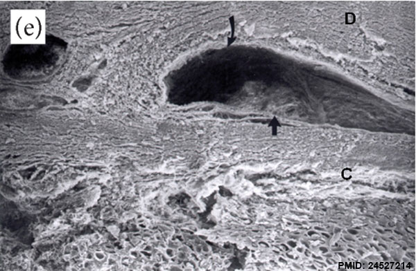
Fetal temporomandibular joint
Human fetus (233 mm GL; 24th week of development). SEM (198).
The inferior joint cavity on the fibrous portion (C) of the condyle, the articular disc (D), and the synovial (arrows) is observed.
- Temporomandibular Joint Links: Image - Week 10 | Image - Week 12 | Image - Week 14 | Image - Week 18 | Image - Week 28 | Image - Week 32 | emporomandibular Joint | Joint Development | Skull Development | Head Development
Reference
<pubmed>24527214</pubmed>| Anat Res Int.
Copyright
© 2014 Carlos Sabu Alvez et al. This is an open access article distributed under the Creative Commons Attribution License, which permits unrestricted use, distribution, and reproduction in any medium, provided the original work is properly cited.
Figure 1: (e) 732720.fig.001e.jpg adjusted in size, labelling and sharpness.
File history
Click on a date/time to view the file as it appeared at that time.
| Date/Time | Thumbnail | Dimensions | User | Comment | |
|---|---|---|---|---|---|
| current | 10:11, 18 February 2015 | | 600 × 392 (71 KB) | Z8600021 (talk | contribs) |
You cannot overwrite this file.
File usage
The following page uses this file: