File:Fetal temporomandibular joint 04.jpg
Fetal_temporomandibular_joint_04.jpg (600 × 390 pixels, file size: 67 KB, MIME type: image/jpeg)
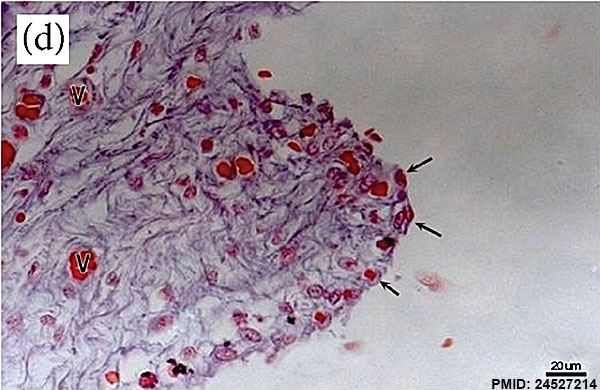
Fetal Temporomandibular Joint
| Human fetus (175 mm GL; 18th week of development). Section of the posterior region of the superior joint cavity. Blood vessel (V); lining cells are visible at the synovial (arrows).
Sagittal section. Trichome Nylceo Marques de Castro. Bar = 200 u m.
|
|
- Temporomandibular Joint Links: Image - Week 10 | Image - Week 12 | Image - Week 14 | Image - Week 18 | Image - Week 28 | Image - Week 32 | emporomandibular Joint | Joint Development | Skull Development | Head Development
Reference
Alvez CS, Carvalho de Moraes LO, Marques SR, Tedesco RC, Harb LJ, Rodríguez-Vázquez JF, Mérida-Velasco JR & Alonso LG. (2014). Analysis by Light, Scanning, and Transmission Microscopy of the Intima Synovial of the Temporomandibular Joint of Human Fetuses during the Development. Anat Res Int , 2014, 732720. PMID: 24527214 DOI.
Copyright
© 2014 Carlos Sabu Alvez et al. This is an open access article distributed under the Creative Commons Attribution License, which permits unrestricted use, distribution, and reproduction in any medium, provided the original work is properly cited.
Figure 1: (d) 732720.fig.001d.jpg adjusted in size, labelling and sharpness.
Cite this page: Hill, M.A. (2024, April 27) Embryology Fetal temporomandibular joint 04.jpg. Retrieved from https://embryology.med.unsw.edu.au/embryology/index.php/File:Fetal_temporomandibular_joint_04.jpg
- © Dr Mark Hill 2024, UNSW Embryology ISBN: 978 0 7334 2609 4 - UNSW CRICOS Provider Code No. 00098G
File history
Click on a date/time to view the file as it appeared at that time.
| Date/Time | Thumbnail | Dimensions | User | Comment | |
|---|---|---|---|---|---|
| current | 10:04, 18 February 2015 | | 600 × 390 (67 KB) | Z8600021 (talk | contribs) | ==Fetal Temporomandibular Joint== {| | width=500px|Human fetus (125 mm GL; 14th week of development). Sagittal section. Hematoxilyn-eosin. Blood vessel (V) and blood cells (arrows) are visible at the synovial. Sagittal section. {{HE}} Bar = 20u m.... |
You cannot overwrite this file.
File usage
The following page uses this file: