File:Fetal mouth and palate day 64.jpg: Difference between revisions
From Embryology
mNo edit summary |
|||
| Line 2: | Line 2: | ||
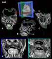
3D scan of the early fetal head week 9 -10 showing the structure of the oral cavity, palate and nasal cavity development. | 3D scan of the early fetal head week 9 -10 showing the structure of the oral cavity, palate and nasal cavity development. | ||
Phosphotungstic acid ( | Phosphotungstic acid (PTAH) staining followed by Computed Tomography (CT) scanning. | ||
:'''Links:''' [[Palate Development]] | [[Fetal Development]] | :'''Links:''' [[Palate Development]] | [[Fetal Development]] | ||
Latest revision as of 09:24, 21 February 2017
Human Fetal Palate (day 64)
3D scan of the early fetal head week 9 -10 showing the structure of the oral cavity, palate and nasal cavity development.
Phosphotungstic acid (PTAH) staining followed by Computed Tomography (CT) scanning.
- Links: Palate Development | Fetal Development
Reference
Image contributed by Contributors#Dr_Joy_Richman Dr Joy Richman.
Original file name - Fig.4_PTA.v2.jpg
Cite this page: Hill, M.A. (2024, May 19) Embryology Fetal mouth and palate day 64.jpg. Retrieved from https://embryology.med.unsw.edu.au/embryology/index.php/File:Fetal_mouth_and_palate_day_64.jpg
- © Dr Mark Hill 2024, UNSW Embryology ISBN: 978 0 7334 2609 4 - UNSW CRICOS Provider Code No. 00098G
File history
Click on a date/time to view the file as it appeared at that time.
| Date/Time | Thumbnail | Dimensions | User | Comment | |
|---|---|---|---|---|---|
| current | 09:21, 21 February 2017 |  | 1,890 × 2,126 (306 KB) | Z8600021 (talk | contribs) | ==Human Fetal Palate (day 64)== 3D scan of the early fetal head week 9 -10 showing the structure of the oral cavity, palate and nasal cavity development. ===Reference=== Image contributed by Contributors#Dr_Joy_Richman Dr Joy Richman. Original... |
You cannot overwrite this file.
File usage
There are no pages that use this file.