File:Fetal ductus venosus ultrasound 01.jpg: Difference between revisions
From Embryology
mNo edit summary |
mNo edit summary |
||
| Line 4: | Line 4: | ||
:'''Links:''' [[:File:Fetal ductus venosus ultrasound 01.jpg|Image - ductus venosus ultrasound]] | [[:File:Fetal_ductus_venosus_pressure_wave_01.jpg|Image - ductus venosus pressure wave]] | [[Ultrasound]] | :'''Links:''' [[:File:Fetal ductus venosus ultrasound 01.jpg|Image - ductus venosus ultrasound]] | [[:File:Fetal_ductus_venosus_pressure_wave_01.jpg|Image - ductus venosus pressure wave]] | [[:File:Gray0502.jpg|Image - fetal circulation]] | [[Ultrasound]] | ||
===Reference=== | ===Reference=== | ||
Revision as of 10:00, 23 August 2014
Fetal ductus venous ultrasound
Human fetal (GA 25 to 32 weeks) ultrasound to show ductus venous (white ring).
- Links: Image - ductus venosus ultrasound | Image - ductus venosus pressure wave | Image - fetal circulation | Ultrasound
Reference
<pubmed>17374167</pubmed>| Cardiovasc Ultrasound.
Copyright
© 2007 da Silva et al; licensee BioMed Central Ltd. This is an Open Access article distributed under the terms of the Creative Commons Attribution License (http://creativecommons.org/licenses/by/2.0), which permits unrestricted use, distribution, and reproduction in any medium, provided the original work is properly cited.
File history
Click on a date/time to view the file as it appeared at that time.
| Date/Time | Thumbnail | Dimensions | User | Comment | |
|---|---|---|---|---|---|
| current | 18:47, 22 August 2014 | 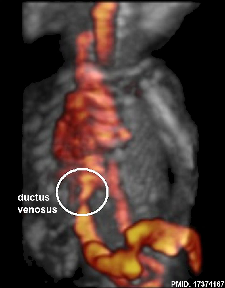 | 783 × 1,000 (68 KB) | Z8600021 (talk | contribs) | ==Fetal ductus venous ultrasound== {{GA}} 25 to 32 weeks |
You cannot overwrite this file.
File usage
The following 3 pages use this file: