File:Fetal ductus venosus ultrasound 01.jpg
Original file (783 × 1,000 pixels, file size: 68 KB, MIME type: image/jpeg)
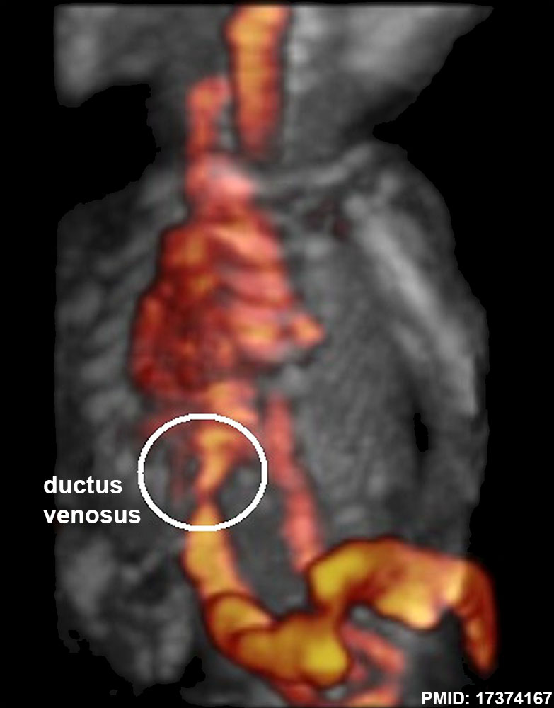
Fetal Ductus Venous Ultrasound
Human fetal (GA 25 to 32 weeks) ultrasound to show ductus venous (white ring). See also cartoon of fetal circulation.
- Ductus Venosus Links: Image - ductus venosus ultrasound | Image - ductus venosus pressure wave | Image - fetal circulation | Ultrasound
Reference
da Silva FC, de Sá RA, de Carvalho PR & Lopes LM. (2007). Doppler and birth weight Z score: predictors for adverse neonatal outcome in severe fetal compromise. Cardiovasc Ultrasound , 5, 15. PMID: 17374167 DOI.
Copyright
© 2007 da Silva et al; licensee BioMed Central Ltd. This is an Open Access article distributed under the terms of the Creative Commons Attribution License (http://creativecommons.org/licenses/by/2.0), which permits unrestricted use, distribution, and reproduction in any medium, provided the original work is properly cited.
Cite this page: Hill, M.A. (2024, April 27) Embryology Fetal ductus venosus ultrasound 01.jpg. Retrieved from https://embryology.med.unsw.edu.au/embryology/index.php/File:Fetal_ductus_venosus_ultrasound_01.jpg
- © Dr Mark Hill 2024, UNSW Embryology ISBN: 978 0 7334 2609 4 - UNSW CRICOS Provider Code No. 00098G
File history
Click on a date/time to view the file as it appeared at that time.
| Date/Time | Thumbnail | Dimensions | User | Comment | |
|---|---|---|---|---|---|
| current | 18:47, 22 August 2014 | | 783 × 1,000 (68 KB) | Z8600021 (talk | contribs) | ==Fetal ductus venous ultrasound== {{GA}} 25 to 32 weeks |
You cannot overwrite this file.
File usage
The following 3 pages use this file: