File:Fetal cardiac state diagram 01.jpg
From Embryology
Size of this preview: 800 × 214 pixels. Other resolution: 1,200 × 321 pixels.
Original file (1,200 × 321 pixels, file size: 29 KB, MIME type: image/jpeg)
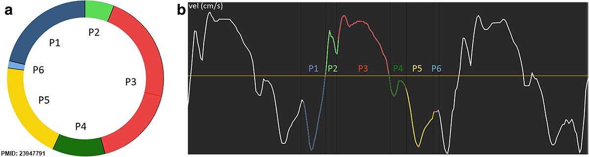
Fetal Cardiac State Diagram (CSD)
- a - a cardiac state diagram CSD of septum from a healthy fetus at 36 weeks of gestation
- b - the extracted myocardial velocity (thick curve) and acceleration (thin curve) from which the CSD was generated.
Reference
<pubmed>23947791</pubmed>| Cardiovasc Ultrasound.
Copyright
© 2013 Elmstedt et al.; licensee BioMed Central Ltd. This is an Open Access article distributed under the terms of the Creative Commons Attribution License (http://creativecommons.org/licenses/by/2.0), which permits unrestricted use, distribution, and reproduction in any medium, provided the original work is properly cited.
Figure 1. Adjusted in size and labelling.
File history
Click on a date/time to view the file as it appeared at that time.
| Date/Time | Thumbnail | Dimensions | User | Comment | |
|---|---|---|---|---|---|
| current | 11:20, 28 August 2014 | 1,200 × 321 (29 KB) | Z8600021 (talk | contribs) | ==Fetal cardiac state diagram== The cardiac state diagram (CSD). Example displaying (a) a cardiac state diagram CSD of septum from a healthy fetus at 36 weeks of gestation, and (b) the extracted myocardial velocity (thick curve) and acceleration (thi... |
You cannot overwrite this file.
File usage
There are no pages that use this file.