File:Fawcett 1910 fig03.jpg: Difference between revisions
From Embryology
({{Fawcett1910_sphenoid_figures}}) |
mNo edit summary |
||
| Line 1: | Line 1: | ||
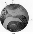
==Fig. 3 Photograph of a sagittal section of the head of the 21 mm embryo== | |||
From which the reconstruction ([[:File:Fawcett 1910 fig02.jpg|fig. 2]]) was made, and at S.R. shows this independent rod of cartilage, which is rounded in section and connected by a fibrous band with the clivus region. In a 30 mm. embryo lent by Professor Bryce of Glasgow this rod of cartilage is no longer independent. | |||
{{Fawcett1910_sphenoid_figures}} | {{Fawcett1910_sphenoid_figures}} | ||
Latest revision as of 09:13, 29 December 2014
Fig. 3 Photograph of a sagittal section of the head of the 21 mm embryo
From which the reconstruction (fig. 2) was made, and at S.R. shows this independent rod of cartilage, which is rounded in section and connected by a fibrous band with the clivus region. In a 30 mm. embryo lent by Professor Bryce of Glasgow this rod of cartilage is no longer independent.
| Historic Disclaimer - information about historic embryology pages |
|---|
| Pages where the terms "Historic" (textbooks, papers, people, recommendations) appear on this site, and sections within pages where this disclaimer appears, indicate that the content and scientific understanding are specific to the time of publication. This means that while some scientific descriptions are still accurate, the terminology and interpretation of the developmental mechanisms reflect the understanding at the time of original publication and those of the preceding periods, these terms, interpretations and recommendations may not reflect our current scientific understanding. (More? Embryology History | Historic Embryology Papers) |
|
|
Reference
Fawcett E. Notes on the development of the human sphenoid. (1910) J Anat. Physiol. 44(3): 207-22. PMID 17232842
Cite this page: Hill, M.A. (2024, May 20) Embryology Fawcett 1910 fig03.jpg. Retrieved from https://embryology.med.unsw.edu.au/embryology/index.php/File:Fawcett_1910_fig03.jpg
- © Dr Mark Hill 2024, UNSW Embryology ISBN: 978 0 7334 2609 4 - UNSW CRICOS Provider Code No. 00098G
File history
Click on a date/time to view the file as it appeared at that time.
| Date/Time | Thumbnail | Dimensions | User | Comment | |
|---|---|---|---|---|---|
| current | 08:31, 29 December 2014 |  | 970 × 996 (202 KB) | Z8600021 (talk | contribs) | {{Fawcett1910_sphenoid_figures}} |
You cannot overwrite this file.
File usage
The following page uses this file:
