File:Fawcett1975 fig34.jpg
Original file (1,280 × 1,746 pixels, file size: 506 KB, MIME type: image/jpeg)
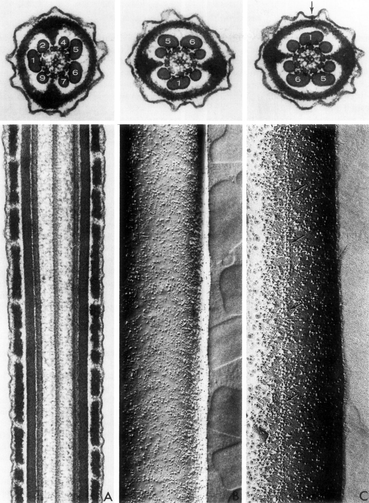
Fig. 34. Spermatozoa Tail (EM)
A - A longitudinal section through the principal piece of a sperm tail, approximally in the plane passing through fibers 2 and 7 or 9 and 4 of the cross-section shown above. The section includes two doublets and one of the central pair of axonemal microtubules, two outer fibers and groups of partially fused ribs of the fibrous sheath.
B - A freeze cleaving preparation of the membrane on the side of the principal piece over the major compartment containing outer fibers 4 to 7. There is a high concentration of randomly distributed membrane intercalated particles.
C - A freeze-cleave preparation of the membrane on the side of the minor compartment containing fibers 9, 1 and 2. A double row of large particles runs longitudinally within the membrane overlying fiber number one. A slight thickening of the membrane at this site is evident (at the arrow) in the cross-section shown above.
- Links: Spermatozoa Development
Reference
From D. W. Fawcett, D. S. Friend, M. Price and R. W. Linck, Proc. 8th Intemat. Congr. Electron Microscopy, Canberra, 1974
Fawcett DW. The Mammalian Spermatozoon. (1975) Dev. Biol. 44, 394-436.
Cite this page: Hill, M.A. (2024, April 28) Embryology Fawcett1975 fig34.jpg. Retrieved from https://embryology.med.unsw.edu.au/embryology/index.php/File:Fawcett1975_fig34.jpg
- © Dr Mark Hill 2024, UNSW Embryology ISBN: 978 0 7334 2609 4 - UNSW CRICOS Provider Code No. 00098G
File history
Click on a date/time to view the file as it appeared at that time.
| Date/Time | Thumbnail | Dimensions | User | Comment | |
|---|---|---|---|---|---|
| current | 09:54, 20 August 2017 | | 1,280 × 1,746 (506 KB) | Z8600021 (talk | contribs) | |
| 09:53, 20 August 2017 | 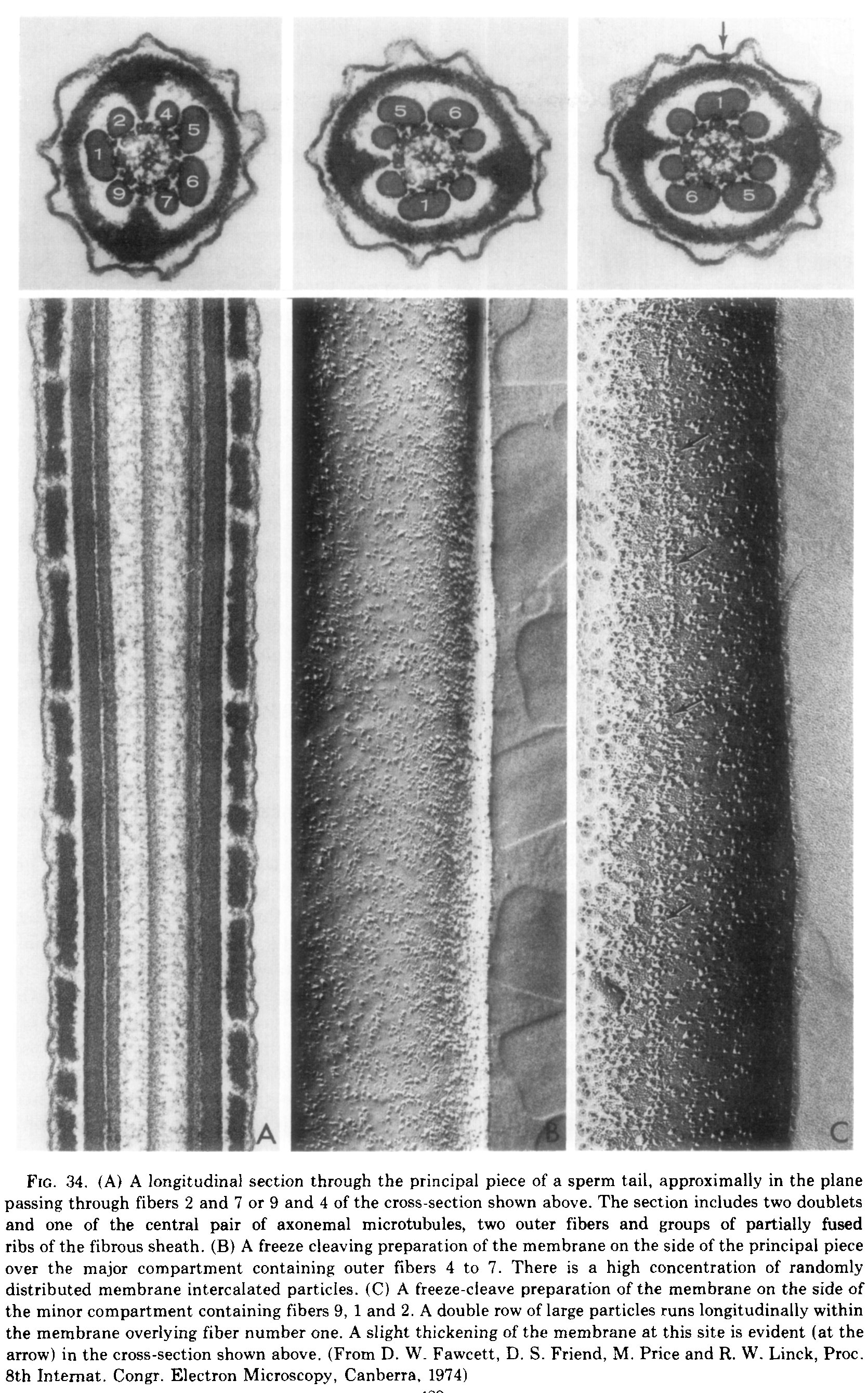 | 1,670 × 2,679 (838 KB) | Z8600021 (talk | contribs) | ==Fig. 34.== ===Reference=== {{Ref-Fawcett1975}} {{Footer}} |
You cannot overwrite this file.
File usage
The following page uses this file: