File:Fawcett1975 fig31.jpg
From Embryology

Size of this preview: 800 × 252 pixels. Other resolution: 1,280 × 403 pixels.
Original file (1,280 × 403 pixels, file size: 128 KB, MIME type: image/jpeg)
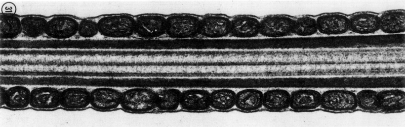
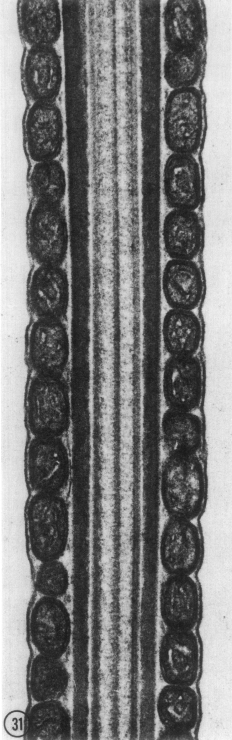
Fig. 31. A longitudinal thin section of the middle piece of a mammalian spermatozoon
The circumferentially oriented mitochondria are cut transversely. Note how closely the celi membrane is apposed to the underlying mitochondria.
- Links: Spermatozoa Development
Reference
From D. W. Fawcett, D. S. Friend, M. Price and R. W. Linck, Proc. 8th Intemat. Congr. Electron Microscopy, Canberra, 1974
Fawcett DW. The Mammalian Spermatozoon. (1975) Dev. Biol. 44, 394-436.
Cite this page: Hill, M.A. (2024, April 28) Embryology Fawcett1975 fig31.jpg. Retrieved from https://embryology.med.unsw.edu.au/embryology/index.php/File:Fawcett1975_fig31.jpg
- © Dr Mark Hill 2024, UNSW Embryology ISBN: 978 0 7334 2609 4 - UNSW CRICOS Provider Code No. 00098G
File history
Click on a date/time to view the file as it appeared at that time.
| Date/Time | Thumbnail | Dimensions | User | Comment | |
|---|---|---|---|---|---|
| current | 10:02, 20 August 2017 | 1,280 × 403 (128 KB) | Z8600021 (talk | contribs) | ||
| 10:02, 20 August 2017 | 740 × 2,353 (269 KB) | Z8600021 (talk | contribs) | FIG. 31. A longitudinal thin section of the middle piece of a mammalian spermatozoon. The circumferen- tially oriented mitochondria are cut transversely. Note how closely the celi membrane is apposed to the underlying mitochondria. |
You cannot overwrite this file.
File usage
The following page uses this file: