File:Eye and retina cartoon.jpg: Difference between revisions
From Embryology
No edit summary |
No edit summary |
||
| Line 1: | Line 1: | ||
==Eye and Retina== | |||
{| | |||
| valign="top"|'''The Eye''' | |||
* An enlarged diagram of the fovea is shown in the box. | |||
* Retina forms the inner lining of the most of the posterior part of the eye. | |||
* The RPE is sandwiched between the retina and choroids, a vascularized and pigmented connective tissue. | |||
| valign="top"|'''Retinal Cell Organization''' | |||
R, rod; C, cone; B, bipolar cell; H, horizontal cell; A, amacrine cell; G, ganglion cells; M, Müller cell. | |||
| valign="top"|'''Human Retina Histology''' | |||
* An H&E stained transverse section of human retina laminated layers. | |||
* The nuclei of the photoreceptors constitute the outer nuclear layer (ONL). | |||
* The nuclei of the bipolar cells, amacrine cells, horizontal cells, and Müller glial cells are found in the inner nuclear layer (INL), and the nuclei of ganglion cells form the ganglion cell layer (GCL). | |||
* The outer plexiform layer (OPL) contains the processes and synaptic terminals of photoreceptors, horizontal cells, and bipolar cells. | |||
* The inner plexiform layer (IPL) contains the processes and terminals of bipolar cells, amacrine cells, and ganglion cells. | |||
* The processes of Müller glial cells fill all space in the retina that is not occupied by neurons and blood vessels. | |||
|} | |||
Retina histology image - Swaroop and Zack (2002), published by BioMed Central. | |||
Revision as of 14:55, 4 March 2012
Eye and Retina
The Eye
|
Retinal Cell Organization
R, rod; C, cone; B, bipolar cell; H, horizontal cell; A, amacrine cell; G, ganglion cells; M, Müller cell. |
Human Retina Histology
|
Retina histology image - Swaroop and Zack (2002), published by BioMed Central.
File history
Click on a date/time to view the file as it appeared at that time.
| Date/Time | Thumbnail | Dimensions | User | Comment | |
|---|---|---|---|---|---|
| current | 14:50, 4 March 2012 | 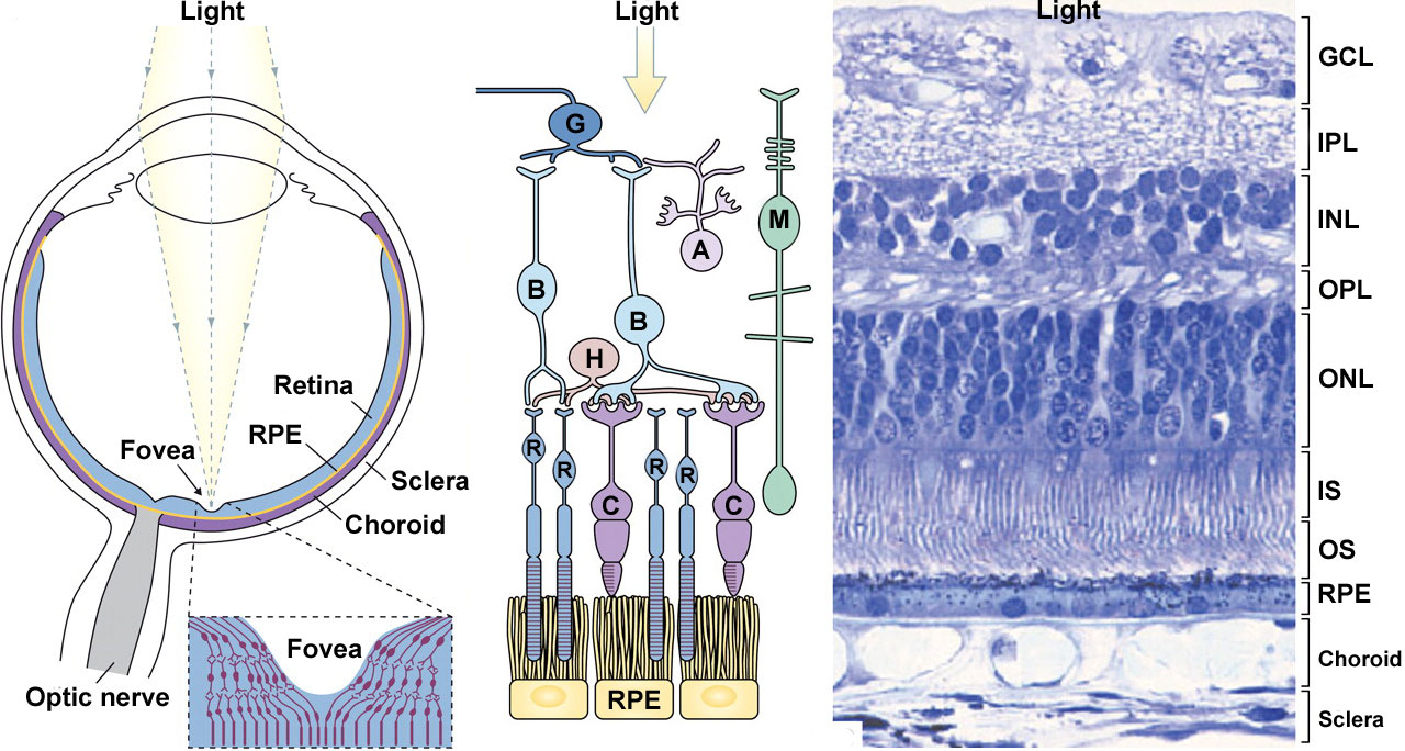 | 1,280 × 684 (211 KB) | Z8600021 (talk | contribs) |
You cannot overwrite this file.