File:Eye-neural crest signaling.jpg
Original file (946 × 886 pixels, file size: 436 KB, MIME type: image/jpeg)
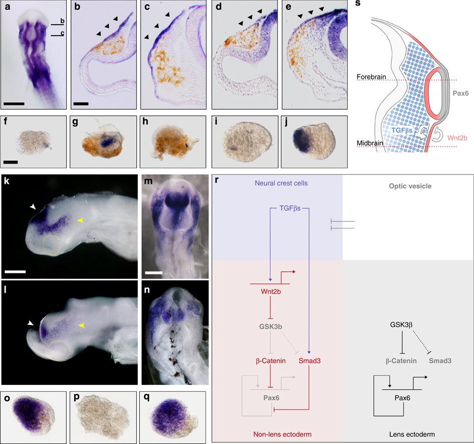
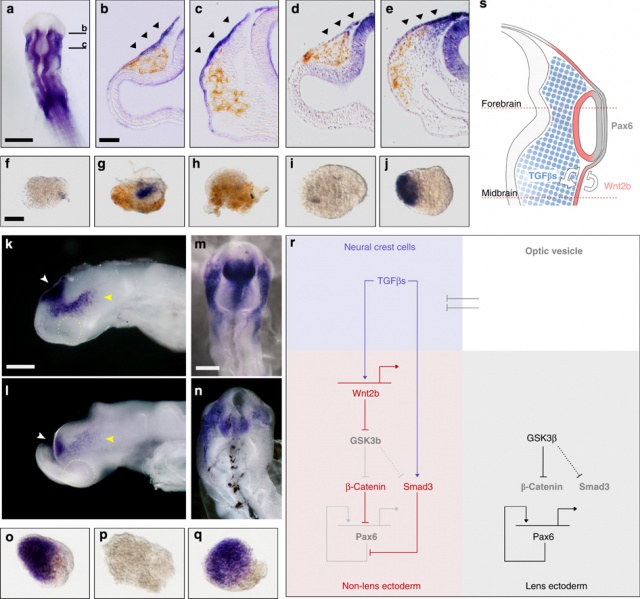
Wnt mediates lens repression by neural crest cells and Transforming growth factor-β
- Transforming growth factor β (TGF-β)
- Neural crest cell (NCC)
(a–c) Wnt2b (blue) gene expression during neural crest cell (NCC) migration:
(a) Whole-mount embryo showing the forebrain section used in panel b and the midbrains section used in panel c. (b) Forebrain level section stained for Wnt2b; (c) midbrain level section stained for Wnt2b.
(d) Forebrain level section stained for Axin2 gene expression (blue). (e) Midbrain level section showing Axin2 expression. All sections are immunostained for NCC-specific HNK1 (brown). Arrowheads indicate gene expression in non-lens ectoderm.
(f–h) Presumtive lens ectoderm (PLE) explants were cultured alone or in combination with NCC or SB431542 and assayed for Wnt2b gene expression (blue) and HNK1 (brown): (f) PLE alone; (g) PLE+NCC; (h) PLE+NCC+SB431542.
(i, j) PLE explants were cultured alone or with Activin A and assayed for Wnt2b gene expression: (i) PLE alone; (j) PLE+Activin A.
(k, l) Wnt2b gene expression (blue) following in ovo ablation of premigratory NCCs: (k) control embryo; (l) NCC ablated embryo. Compare Wnt2b expression in the brain (white arrowheads) with non-lens surface ectoderm (yellow arrowheads).
(m, n) Wnt2b gene expression (blue) following in ovo electroporation of expression vectors encoding green fluorescent protein (GFP) or Smad7+GFP: (m) GFP; (n) Smad7+GFP.
(o–q) PLE explants were cultured alone or in combination with Activin A or N-Fz8 and assayed for Pax6 gene expression (blue): (o) PLE alone; (p) PLE+Activin A; (q) PLE+Activin A+N-Fz8.
(r) Proposed molecular model to explain TGF-β- and Wnt-mediated lens restriction. Broken lines: interactions inferred from the literature.
(s) Proposed embryological model summarizing how NCCs organize the eye: NCCs (blue) secrete TGF-βs, which signal to the non-lens ectoderm and dorsal optic vesicle. As a consequence, Wnt2b (red) is induced, and together they repress lens formation in the non-lens ectoderm. This leads to the alignment of Pax6 expression in the future lens and neural retina (grey).
Scale bars: a, 500 μm; b, 50 μm for panels b–e; f, 50 μm for panels f–j, o–q; k, 250 μm for panels k, l; m, 250 μm for panels m, n.
- Links: Lens Development | Lens repression by neural crest cells | Proposed model how NCCs organize the eye | molecular model to explain TGF-β- and Wnt-mediated lens restriction
Original image name: Figure 3 http://www.nature.com/ncomms/journal/v2/n4/fig_tab/ncomms1269_F3.html
Reference
Grocott T, Johnson S, Bailey AP, Streit A. Neural crest cells organize the eye via TGF-β and canonical Wnt signalling. Nat Commun. 2011 Apr;2:265. PMID21468017 | Nat Commun.
The request you have made is considered to be non-commercial/educational. As the article you have requested has been distributed under a Creative Commons license (Attribution-Noncommercial 2.5), you may reuse this material for non-commercial/educational purposes without obtaining additional permission from Nature Publishing Group, providing that the author and the original source of publication are fully acknowledged.
For full terms and conditions of the Creative Commons license, please see the attached link http://creativecommons.org/licenses/by-nc/2.5
File history
Click on a date/time to view the file as it appeared at that time.
| Date/Time | Thumbnail | Dimensions | User | Comment | |
|---|---|---|---|---|---|
| current | 13:51, 29 April 2011 | | 946 × 886 (436 KB) | S8600021 (talk | contribs) | ==Wnt mediates lens repression by neural crest cells and Transforming growth factor-β== * Transforming growth factor β (TGF-β) * Neural crest cell (NCC) (a–c) Wnt2b (blue) gene expression during neural crest cell (NCC) migration: (a) Whole-mount |
You cannot overwrite this file.
File usage
The following page uses this file: