File:Ewart1897 07.jpg

Original file (1,200 × 757 pixels, file size: 167 KB, MIME type: image/jpeg)
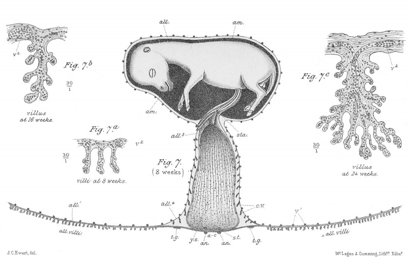
Fig. 7. Eight-weeks (56 days) Horse Embryo
Represents an eight-weeks' (56 days) horse embryo suspended in the amniotic cavity, and bathed by the amniotic fluid. The embryo is connected with the yolk sac (y.s.) and the allantois (only part of which is shown) by relatively large blood-vessels. These vessels and their investing tissues form the proximal part of the umbilical cord (sta.). The yolk sac, though having still as great a capacity as formerly, is folded longitudinally, so as to occupy a comparatively small space. Note that the absorbing area is now very small, and especially that numerous simple villi now project from the surface of the embryonic sac. Each villus consists of a vascular allantoic core, and a thin capsule derived from the embryonic sac. The villi are represented on too large a scale, but the embryo and yolk sac are natural size.
7a . Represents three villi from an eight weeks' embryo.
7b. A single villus from a sixteen-weeks' embryo, and
7c. A single villus from a twenty-four-weeks' embryo — all thirty times natural size.
The villi fit into pits or moulds in the lining of the uterus. As the foetal blood circulates through the villi it comes sufficiently near the maternal blood circulating through the wall of the complex pits or moulds to admit of an exchange of fluids and gases— the foetal blood transfers its excess of carbonic acid to the maternal blood, while the maternal transfers to the foetal blood some of its oxygen, and also fluids containing all the ingredients required for the development and growth of the embryo. There is, however, no actual mixing of the maternal and foetal blood. At birth the foetal villi are simply withdrawn from the uterine pits. The villi and pits together are generally spoken of as the Placenta.
| Historic Disclaimer - information about historic embryology pages |
|---|
 |
Reference
Ewart, J.C. A Critical Period in the Development of the Horse. London: Adam and Charles Black (1897).
File history
Click on a date/time to view the file as it appeared at that time.
| Date/Time | Thumbnail | Dimensions | User | Comment | |
|---|---|---|---|---|---|
| current | 15:45, 3 May 2013 | | 1,200 × 757 (167 KB) | Z8600021 (talk | contribs) | {{Ewart1897 figures}} |
You cannot overwrite this file.
File usage
The following page uses this file: