File:Ewart1897 06.jpg

Original file (1,200 × 771 pixels, file size: 93 KB, MIME type: image/jpeg)
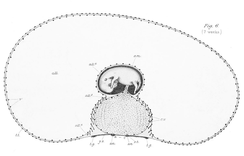
Fig. 6. The seven-weeks (49 days) Horse Embryo
Contrast this with the other embryos, more especially with the five and eight weeks' embryos. Note that while the yolk sac has remained almost stationary, the absorbing area has diminished, while the capacity of the allantois (all.) has greatly increased. The villi (c.v.) extending from the allantois towards the yolk sac are numerous, long and slender, but still devoid of blood-vessels. Indications of the coming external vascular villi occur in the form of minute dots (t.t.) over the surface of the embryonic sac. The yolksac and allantoic stalks have already united at their inner or proximal ends.
| Historic Disclaimer - information about historic embryology pages |
|---|
 |
Reference
Ewart, J.C. A Critical Period in the Development of the Horse. London: Adam and Charles Black (1897).
File history
Click on a date/time to view the file as it appeared at that time.
| Date/Time | Thumbnail | Dimensions | User | Comment | |
|---|---|---|---|---|---|
| current | 15:54, 3 May 2013 | | 1,200 × 771 (93 KB) | Z8600021 (talk | contribs) |
You cannot overwrite this file.
File usage
The following page uses this file: