File:Evatt1909 fig01-04.jpg

Original file (1,000 × 1,137 pixels, file size: 169 KB, MIME type: image/jpeg)
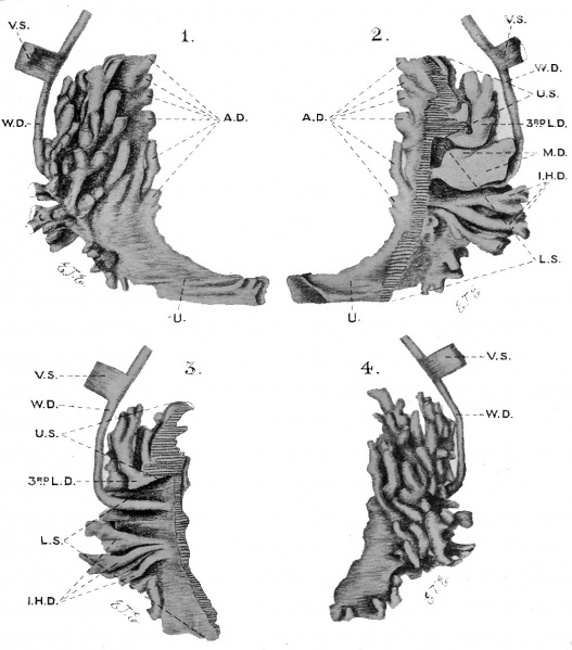
Fig. 1 to 4
Different views of the two halves of a wax-plate model of the rostatic urethra and glandular tissue of the prostate. The short transverse lines in figs. 2 and 3 in icate the plane along which the model was split.
Figs. 1 and 2, outer and inner surfaces of the right half respectively. Figs. 3 and 4, inner and outer surfaces of the left half respectively. U.S., up r subdivision ; L.S., lower subdivision: U., urethra; A.D., anterior ducts ; I.lI.D., inferior horizontal ducts; 3r L.D.. ducts of the “3rd lobe ”; W.D., Wolflian ducts; V.S., vesicular seminnles ; M.D., Miillerian ducts.
Reference
Evatt EJ. A contribution to the development of the prostate in man. (1909) Jour. of Anat. and Phys. 43: 314-321.
Cite this page: Hill, M.A. (2024, April 28) Embryology Evatt1909 fig01-04.jpg. Retrieved from https://embryology.med.unsw.edu.au/embryology/index.php/File:Evatt1909_fig01-04.jpg
- © Dr Mark Hill 2024, UNSW Embryology ISBN: 978 0 7334 2609 4 - UNSW CRICOS Provider Code No. 00098G
File history
Click on a date/time to view the file as it appeared at that time.
| Date/Time | Thumbnail | Dimensions | User | Comment | |
|---|---|---|---|---|---|
| current | 13:28, 17 June 2017 | | 1,000 × 1,137 (169 KB) | Z8600021 (talk | contribs) | |
| 13:27, 17 June 2017 | 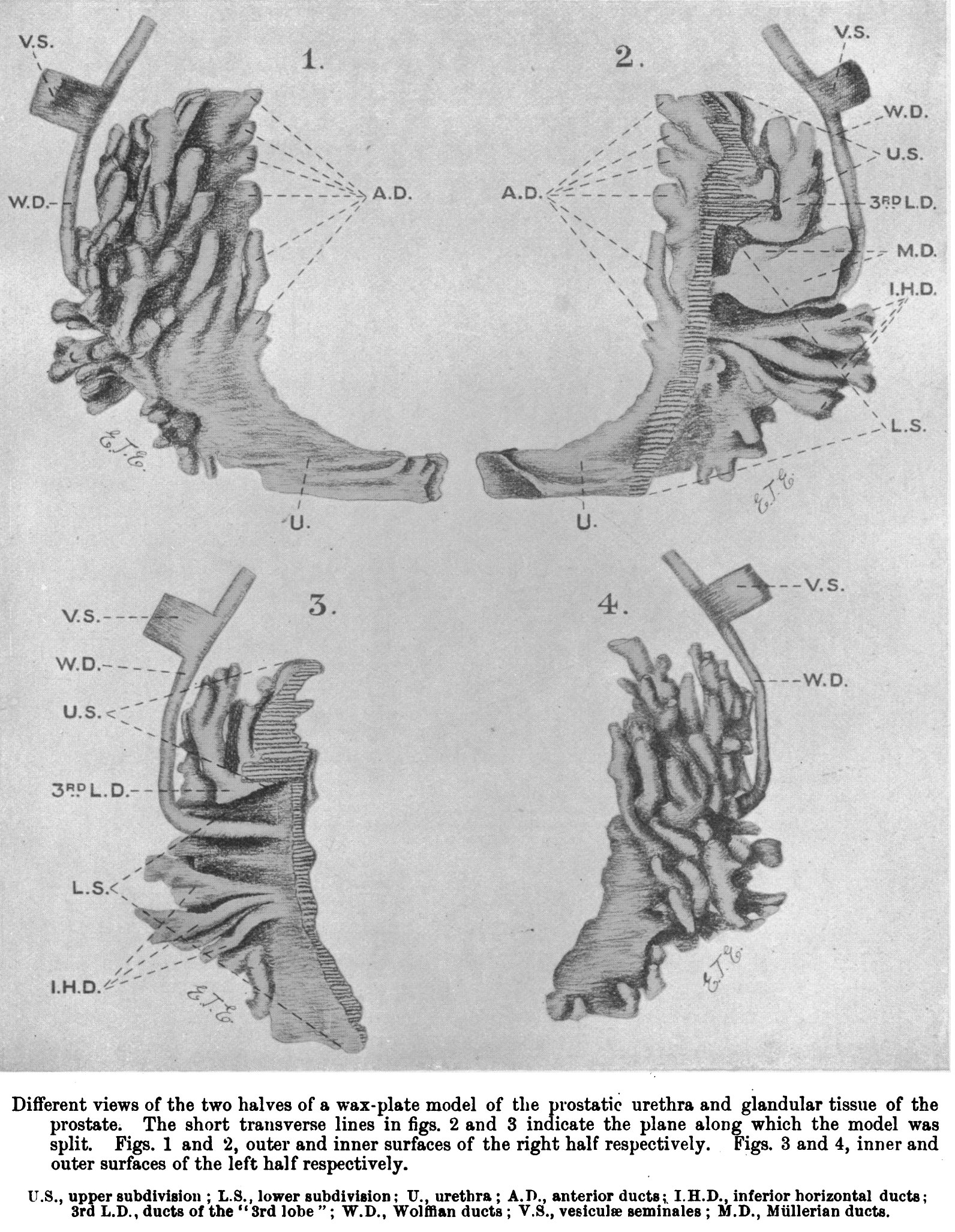 | 1,592 × 2,054 (817 KB) | Z8600021 (talk | contribs) |
You cannot overwrite this file.
File usage
The following page uses this file: