File:Evans1909 fig07-08.jpg

Original file (1,265 × 1,116 pixels, file size: 92 KB, MIME type: image/jpeg)
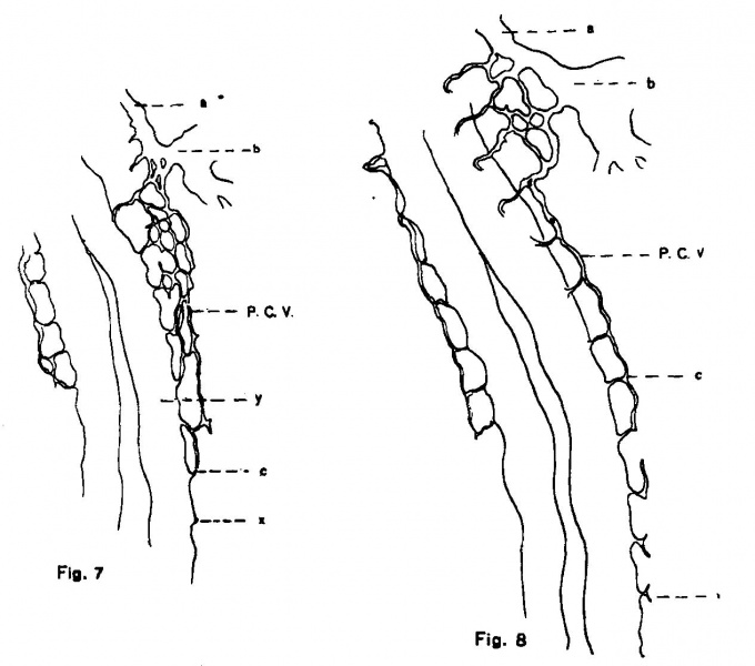
Figs. 7 and 8. Chick embryo of 17 and 21 somites
Fig. 7. View or a total mount of an injected chick embryo of 17 somites, showing the duct of Cuvier and subadjacent region. (X 80.) a = anterior cardinal vein; b = duct of Cuvier; c = 10th segmental vessel; P. C. V. = capillaries from which the poterior cardinal vein is formed; x = endothelial sprout representing the 11th segmental vessel.
Fig. 8. View or a total mount of an injected chick embryo of 21 somites. showing the duct of Cuvier and subjacent region. (x 80.) The lettering is the same as in the preceding figure, with the exception of x, which represents the 14th segmental vessel. One sees the segmental capillaries biturcate often into anterior and posterior sprouts. the union of which makes the continuation of the vein.
| Historic Disclaimer - information about historic embryology pages |
|---|
 |
Evans (1909) Figures: 1 Chick 17 somites | 2 Chick 20 somites | 3 Chick 23 somites | 6 Chick lateral 25 somites |
Reference
Evans HM. On the development of the aortae, cardinal and umbilical veins, and the other blood vessels of vertebrate embryos from capillaries. (1909) Anat. Rec. 3: 498-518.
Cite this page: Hill, M.A. (2024, April 27) Embryology Evans1909 fig07-08.jpg. Retrieved from https://embryology.med.unsw.edu.au/embryology/index.php/File:Evans1909_fig07-08.jpg
- © Dr Mark Hill 2024, UNSW Embryology ISBN: 978 0 7334 2609 4 - UNSW CRICOS Provider Code No. 00098G
File history
Click on a date/time to view the file as it appeared at that time.
| Date/Time | Thumbnail | Dimensions | User | Comment | |
|---|---|---|---|---|---|
| current | 15:57, 28 November 2017 | | 1,265 × 1,116 (92 KB) | Z8600021 (talk | contribs) |
You cannot overwrite this file.