File:Epidermis cartoon 01.jpg: Difference between revisions
| Line 1: | Line 1: | ||
==General structure of the Adult Epidermis== | ==General structure of the Adult Epidermis== | ||
This cartoon shows keratinocyte differentiation within overall structure of the skin epithelium. In addition, the cellular junctions are also indicated. | This cartoon shows keratinocyte differentiation (cornification) within overall structure of the skin epithelium. In addition, the cellular junctions are also indicated. | ||
# '''stratum basale''' - (basal layer) keratinocytes proliferate by mitosis. This layer also contains the stem cell population. | |||
# '''stratum spinosum''' - keratinocytes reinforce their cytoskeletal keratin filament network and adjacent cells interact through desmosomes. | |||
# '''stratum granulosum''' - keratinocytes become more flattened and express proteins that aggregate to form keratohyalin granules. Lipids are also produced and stored in lamellar bodies. | |||
# '''stratum corneum''' - keratinocytes lose their organelles, including the nucleus, and become the dead, flattened corneocytes. | |||
===Reference=== | ===Reference=== | ||
Revision as of 14:39, 17 May 2012
General structure of the Adult Epidermis
This cartoon shows keratinocyte differentiation (cornification) within overall structure of the skin epithelium. In addition, the cellular junctions are also indicated.
- stratum basale - (basal layer) keratinocytes proliferate by mitosis. This layer also contains the stem cell population.
- stratum spinosum - keratinocytes reinforce their cytoskeletal keratin filament network and adjacent cells interact through desmosomes.
- stratum granulosum - keratinocytes become more flattened and express proteins that aggregate to form keratohyalin granules. Lipids are also produced and stored in lamellar bodies.
- stratum corneum - keratinocytes lose their organelles, including the nucleus, and become the dead, flattened corneocytes.
Reference
<pubmed>18250198</pubmed>
J Cell Biol. 2008 February 11; 180(3): 451–458.
doi: 10.1083/jcb.200709098.
Copyright © 2008, The Rockefeller University Press
Copyright
Rockefeller University Press - Copyright Policy This article is distributed under the terms of an Attribution–Noncommercial–Share Alike–No Mirror Sites license for the first six months after the publication date (see http://www.jcb.org/misc/terms.shtml). After six months it is available under a Creative Commons License (Attribution–Noncommercial–Share Alike 4.0 Unported license, as described at https://creativecommons.org/licenses/by-nc-sa/4.0/ ). (More? Help:Copyright Tutorial)
http://www.pubmedcentral.nih.gov/articlerender.fcgi?artid=2234247&rendertype=figure&id=fig1
File history
Click on a date/time to view the file as it appeared at that time.
| Date/Time | Thumbnail | Dimensions | User | Comment | |
|---|---|---|---|---|---|
| current | 14:30, 17 May 2012 | 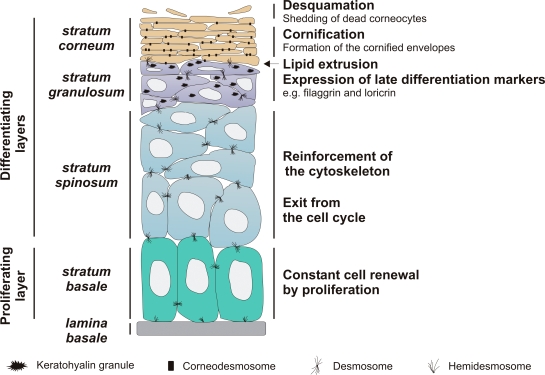 | 545 × 375 (96 KB) | Z8600021 (talk | contribs) | ==General structure of the Adult Epidermis== Introduction excerpt: :"The basal layer, or stratum basale, of the epidermis contains proliferating keratinocytes (Fig. 1). Upon withdrawal from the cell cycle, these basal keratinocytes detach from the basem |
You cannot overwrite this file.
File usage
The following page uses this file: