File:Enbom1939 fig01.jpg

Original file (1,250 × 572 pixels, file size: 55 KB, MIME type: image/jpeg)
Fig.1a and b Diagrammatic side View of plastiline models of opossum embryos, (:1) 9.4 mm. and (b) 11.2 mm. Linear course of the intestinal canal with the omphalomesenteric artery axial in the C-onvolute system. In the older foetus the pliysiological umbilical hernia has been drawn in as a consequence of the vitelline artery having been shortened by one—half, the gastro-enterie coil in its entirety then being pressed together with dorsal tlisplacexnent. A0 = aorta together with a. omphalomesenteriea. C = coecum. S = stomach. ac = point at which vitelline artery leaves mesentery. Large intestine drawn with thicker line. 8/1.
File history
Click on a date/time to view the file as it appeared at that time.
| Date/Time | Thumbnail | Dimensions | User | Comment | |
|---|---|---|---|---|---|
| current | 14:05, 16 February 2018 | | 1,250 × 572 (55 KB) | Z8600021 (talk | contribs) | |
| 14:04, 16 February 2018 | 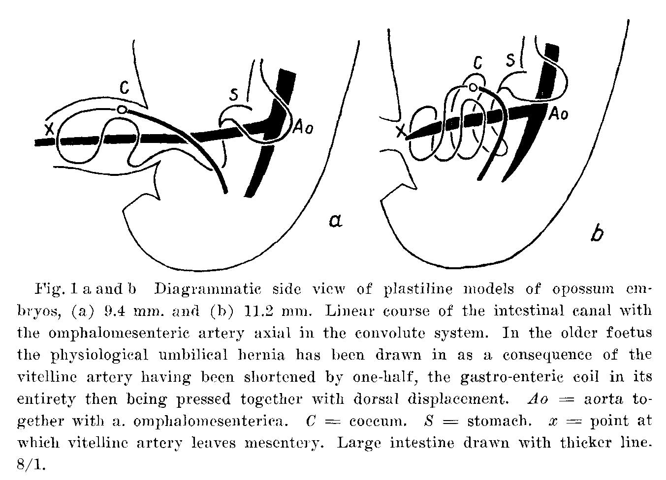 | 1,353 × 977 (155 KB) | Z8600021 (talk | contribs) | '''Fig.1a and b''' Diagrammatic side View of plastiline models of opossum embryos, (:1) 9.4 mm. and (b) 11.2 mm. Linear course of the intestinal canal with the omphalomesenteric artery axial in the C-onvolute system. In the older foetus the pliysiologi... |
You cannot overwrite this file.
File usage
The following page uses this file: