File:Embryonic Circulations.jpg: Difference between revisions
From Embryology
(category:Heart ILP The three early embryonic circulations. Three paired veins drain into the primordial heart tube: vitelline veins (returning poorly oxygenated blood from the yolk sac), umbilical veins (carrying well-oxygenated blood from the primor) |
mNo edit summary |
||
| (2 intermediate revisions by one other user not shown) | |||
| Line 1: | Line 1: | ||
==The Three Early Embryonic Circulations== | |||
Three paired veins drain into the primordial heart tube: | |||
# vitelline veins (returning poorly oxygenated blood from the yolk sac) | |||
# umbilical veins (carrying well-oxygenated blood from the primordial placenta) | |||
# common cardinal veins (return poorly oxygenated blood from the body of the embryo). | |||
The vitelline venous system gives rise to the liver sinusoids and portal system, and forms the ductus venosus which shunts blood from the umbilical vein to the IVC. | |||
{{Intermediate_Cardiac_menu}} | |||
{{Footer}} | |||
[[Category:Heart ILP]] [[Category:Heart]] [[Category:Cardiovascular]] [[Category:Cartoon]] | |||
Latest revision as of 13:06, 26 August 2014
The Three Early Embryonic Circulations
Three paired veins drain into the primordial heart tube:
- vitelline veins (returning poorly oxygenated blood from the yolk sac)
- umbilical veins (carrying well-oxygenated blood from the primordial placenta)
- common cardinal veins (return poorly oxygenated blood from the body of the embryo).
The vitelline venous system gives rise to the liver sinusoids and portal system, and forms the ductus venosus which shunts blood from the umbilical vein to the IVC.
| Begin Intermediate: | Primordial Heart Tube | Heart Tube Looping | Atrial Ventricular Septation | Outflow Tract | Heart Valves | Cardiac Abnormalities | Vascular Overview |
Cite this page: Hill, M.A. (2024, April 26) Embryology Embryonic Circulations.jpg. Retrieved from https://embryology.med.unsw.edu.au/embryology/index.php/File:Embryonic_Circulations.jpg
- © Dr Mark Hill 2024, UNSW Embryology ISBN: 978 0 7334 2609 4 - UNSW CRICOS Provider Code No. 00098G
File history
Click on a date/time to view the file as it appeared at that time.
| Date/Time | Thumbnail | Dimensions | User | Comment | |
|---|---|---|---|---|---|
| current | 10:28, 14 March 2010 | 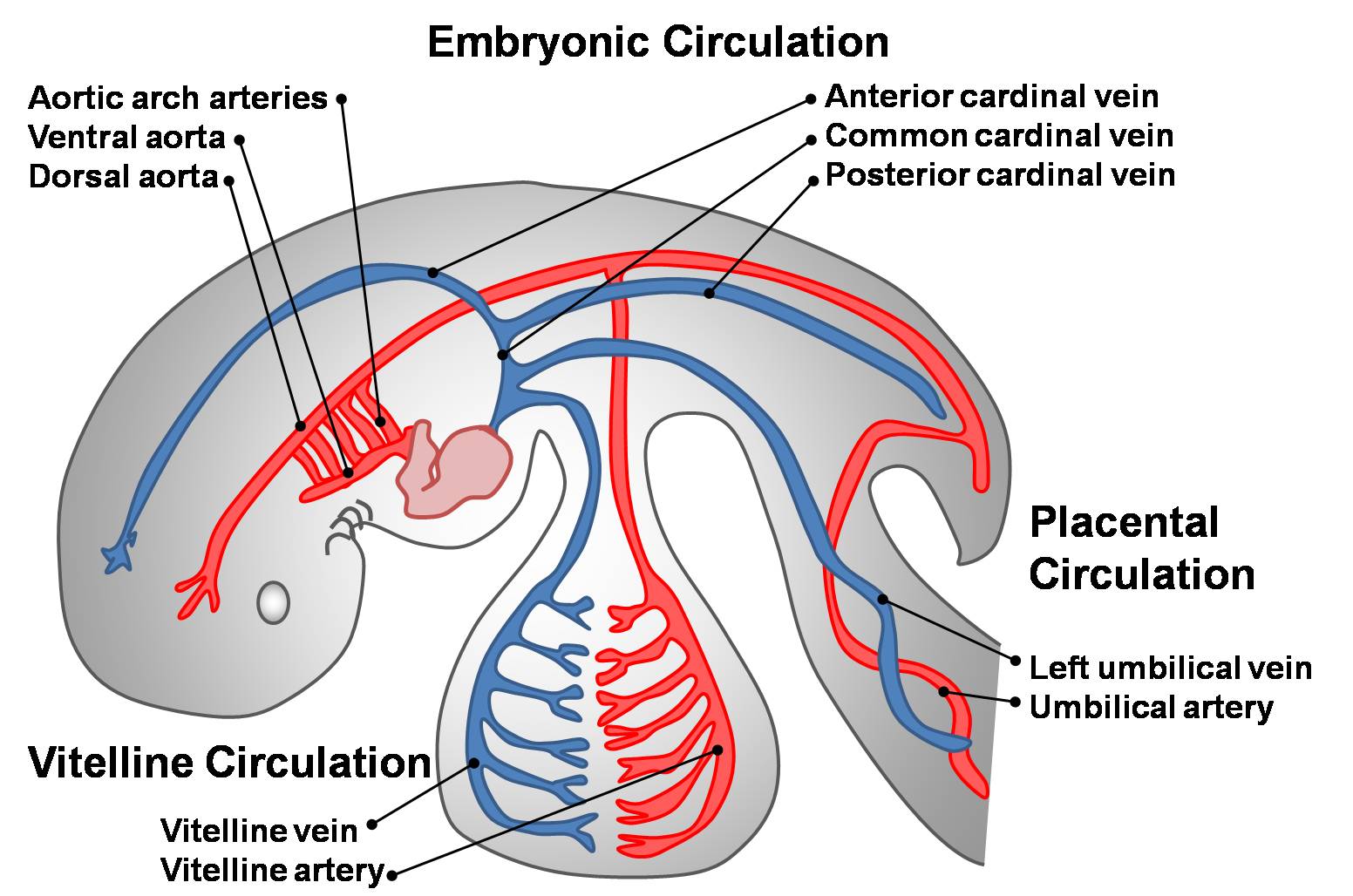 | 1,552 × 1,028 (171 KB) | Z3212774 (talk | contribs) | category:Heart ILP The three early embryonic circulations. Three paired veins drain into the primordial heart tube: vitelline veins (returning poorly oxygenated blood from the yolk sac), umbilical veins (carrying well-oxygenated blood from the primor |
You cannot overwrite this file.