File:Elephant ovary and uterus.jpg

Original file (1,200 × 424 pixels, file size: 61 KB, MIME type: image/jpeg)
Elephant ovary and uterus
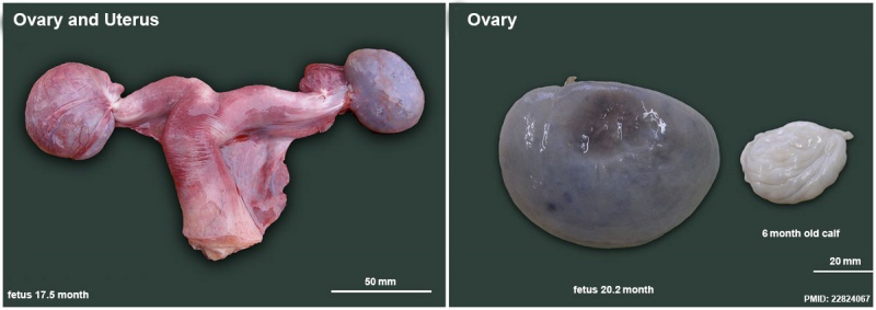
The uterus and ovaries of an elephant fetus at 17.5 month of gestation. The ovary on the left is still enclosed within its ovarial sac (Scale bar = 50 mm).
Ovary from a 20.2 month old elephant fetus on the left and a 6 month old calf on the right showing great shrinkage around the time of birth due to the reduction in both the size and number of interstitial cells (Scale bar = 20 mm).
- Links: Elephant Development
Reference
<pubmed>22824067</pubmed>| BMC Vet Res.
Copyright
© 2012 Stansfield et al.; licensee BioMed Central Ltd. This is an Open Access article distributed under the terms of the Creative Commons Attribution License (http://creativecommons.org/licenses/by/2.0), which permits unrestricted use, distribution, and reproduction in any medium, provided the original work is properly cited.
Figure 5. 1746-6148-8-119-5.jpg Original image adjusted in size and labelling.
File history
Click on a date/time to view the file as it appeared at that time.
| Date/Time | Thumbnail | Dimensions | User | Comment | |
|---|---|---|---|---|---|
| current | 11:01, 7 January 2015 | 1,200 × 424 (61 KB) | Z8600021 (talk | contribs) | ==Elephant ovary and uterus== The uterus and ovaries of an elephant fetus at 17.5 month of gestation. The ovary on the left is still enclosed within its ovarial sac (Scale bar = 50 mm). Ovary from a 20.2 month old elephant fetus on the left... |
You cannot overwrite this file.
File usage
The following page uses this file: