File:Early lung develop.jpg: Difference between revisions
No edit summary |
No edit summary |
||
| (One intermediate revision by the same user not shown) | |||
| Line 7: | Line 7: | ||
Direct journal link: https://www.ncbi.nlm.nih.gov/pmc/articles/PMC5320013/#CR82 | Direct journal link: https://www.ncbi.nlm.nih.gov/pmc/articles/PMC5320013/#CR82 | ||
===Original image source=== | |||
Schittny JC (2014) Strukturelle Entwicklung – von der Anlage zur adulten Lunge Mutius In: von Mutius E, Gappa M, Ebner E, Frey U (eds) Pädiatrische Pneumologie. Springer, Berlin | |||
=== Copyright information === | === Copyright information === | ||
Open Access This article is distributed under the terms of the Creative Commons Attribution 4.0 International License (http://creativecommons.org/licenses/by/4.0/), which permits unrestricted use, distribution, and reproduction in any medium, provided you give appropriate credit to the original author(s) and the source, provide a link to the Creative Commons license, and indicate if changes were made. | Open Access This article is distributed under the terms of the Creative Commons Attribution 4.0 International License (http://creativecommons.org/licenses/by/4.0/), which permits unrestricted use, distribution, and reproduction in any medium, provided you give appropriate credit to the original author(s) and the source, provide a link to the Creative Commons license, and indicate if changes were made. | ||
=== Reference === | |||
Latest revision as of 10:44, 5 October 2017
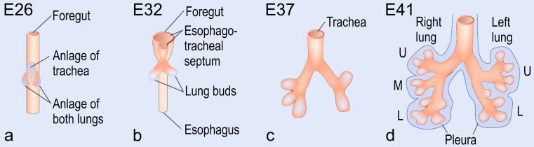
Early lung development
This image demonstrates early human lung development, from the outpouching of lung buds to the formation of the right and left lung.
Original image legend
Early human lung development. At E26, post-conceptional, the anlage of the two lungs forms by outpouchings of the foregut on both sides lateral of the anlage of the trachea (a) (Cardoso and Lu 2006). The prospective trachea forms by a distal-to-proximal segregation from the foregut. At E32, the two lung anlages give rise to the two future main bronchi (b). Due to continued branching, the lobar bronchi are formed at E37 (c). Later, at E41, the segmental bronchi follow (d). Organogenesis is completed after the formation of the pleura (d). U upper lobe; m middle lobe; l lower lobe (from Schittny 2014, by courtesy of Springer, Heidelberg) [1]
Direct journal link: https://www.ncbi.nlm.nih.gov/pmc/articles/PMC5320013/#CR82
Original image source
Schittny JC (2014) Strukturelle Entwicklung – von der Anlage zur adulten Lunge Mutius In: von Mutius E, Gappa M, Ebner E, Frey U (eds) Pädiatrische Pneumologie. Springer, Berlin
Copyright information
Open Access This article is distributed under the terms of the Creative Commons Attribution 4.0 International License (http://creativecommons.org/licenses/by/4.0/), which permits unrestricted use, distribution, and reproduction in any medium, provided you give appropriate credit to the original author(s) and the source, provide a link to the Creative Commons license, and indicate if changes were made.
Reference
- ↑ Schittny, J. C. (2017). Development of the lung. Cell and Tissue Research, 367(3), 427–444. http://doi.org/10.1007/s00441-016-2545-0
File history
Click on a date/time to view the file as it appeared at that time.
| Date/Time | Thumbnail | Dimensions | User | Comment | |
|---|---|---|---|---|---|
| current | 13:00, 4 October 2017 | 761 × 210 (60 KB) | Z5059373 (talk | contribs) |
You cannot overwrite this file.
File usage
The following 2 pages use this file: