File:Duodenal atresia 02.jpg

Original file (765 × 682 pixels, file size: 68 KB, MIME type: image/jpeg)
Summary
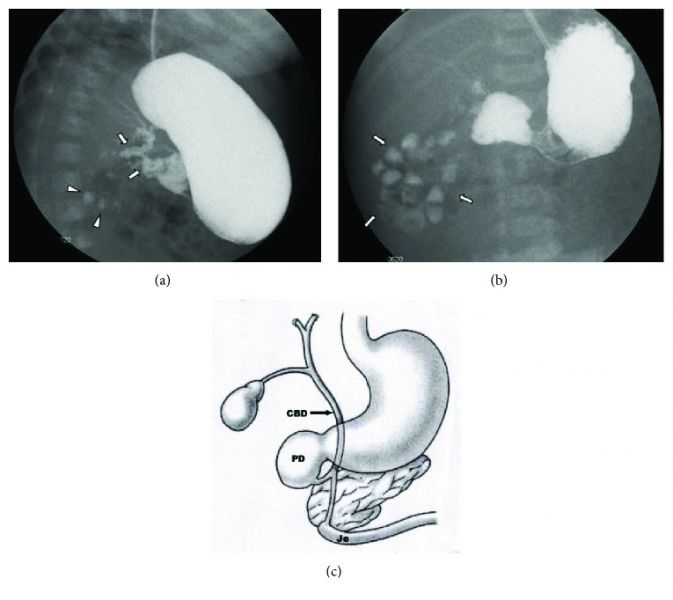
(a) Upper gastrointestinal series showing a complete obstruction of the duodenum and contrast filling of anomalous bifurcated bile ducts (arrows). The small contrast was also noted in the distal bowel (arrowheads). (b) Upper gastrointestinal series showing a complete obstruction at the second portion of the duodenum, and contrast was seen in the proximal jejunum which is located in the right upper quadrant. The proximal location of the jejunum indicates a malrotation of the intestine without evidence of a small bowel obstruction. (c) Diagram showing biliary tract abnormality associated with duodenal atresia (PD—proximal duodenum, Je—jejunum, and CBD—common bile duct).
CRIS2018-8041427.003.jpg
File history
Click on a date/time to view the file as it appeared at that time.
| Date/Time | Thumbnail | Dimensions | User | Comment | |
|---|---|---|---|---|---|
| current | 16:12, 16 April 2019 | | 765 × 682 (68 KB) | Z8600021 (talk | contribs) | (a) Upper gastrointestinal series showing a complete obstruction of the duodenum and contrast filling of anomalous bifurcated bile ducts (arrows). The small contrast was also noted in the distal bowel (arrowheads). (b) Upper gastrointestinal series sho... |
You cannot overwrite this file.
File usage
The following page uses this file: