File:Drosophila and mouse placode similarity.jpg

Original file (499 × 1,086 pixels, file size: 337 KB, MIME type: image/jpeg)
Drosophila and Mouse Placode Similarity
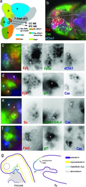
The gt head stripe 1 in stage 10–11 embryos has molecular similarity to the vertebrate hypohyseal placed.
(a) A summary diagram of expression labeling data from b–f, which shows the subdivision of neuroectoderm and underlying neuroblast lineages into 2–12 cell groups with common gene expression. The color-coding for gene expression is given in the diagram. The CC cell and IPC neuroblast progenitors delaminate from the surface neuroectoderm at stages 10 and 12, respectively. At stage 13, regions B (anterior PI) and D (Pars lateralis) invaginate to form neurogenic placodes underneath the surface neuroectoderm and continue to generate neuroblasts (6). The CC cell and IPC neuroblasts are not included in the brain neuroblast map of Urbach and Technau (35) (data not shown). (b–f) All brains labeled by antibodies are as indicated with the text color corresponding to color channels in merged images. Letter labels correspond to those in the diagram (a). Anterior is to the left.
(b) Embryo head with dotted outline showing region of placode gene expression (So, D-six4, Optix, and Eya). The position of the midline is indicated by dotted line. Expression patterns of Eya, Cas, and dChx1.
(c) Expression patterns of Eya, Optix, and dChx1. Region B shows low-level Eya expression.
(d) Expression patterns of Eya, Cas, and gt1. Region F shows high-level gt1 expression.
(e) Expression patterns of So, Cas, and dChx1. The Mz-VUM enhancer of the Dchx1 gene (shown) labels the dChx1+ cells of region A at this stage.
(f) Expression patterns of Fas2, gt1, and Cas. Fas2 labeling of region D corresponds to the developing Pars lateralis (6).
(g) Comparison of hypophyseal gene expression (Optix/Six6 and Eya) in the fate maps of mouse and fly during the early development of the brain endocrine axis and pharynx. The neurohypohyseal diverticulum of the infundibuar region (ir) will give rise to neurosecretory hypothalamic cells (neuhypophysis). Rathke's pouch (rp) is an invagination of the oral ectoderm that gives rise to the anterior pituitary (adenohypohysis). In both mammals and flies, Optix/Six6+ Eya cells are fated to become either pharynx (ph) or endocrine progenitors.
(Scale bar: 5 μm.)
Original file name: Fig. 3. Zpq0490785930003.jpg http://www.ncbi.nlm.nih.gov/pmc/articles/PMC2148390/figure/F3/
Reference
<pubmed>18056636</pubmed>| PMC2148390
Copyright
Proceedings National Academy of Sciences (PNAS) Liberalization of PNAS copyright policy: Noncommercial use freely allowed Note original Author should be contacted for permission to reuse for Educational purposes. See also PNAS Author Rights and Permission FAQs
- Cozzarelli NR, Fulton KR, Sullenberger DM. Liberalization of PNAS copyright policy: noncommercial use freely allowed. Proc Natl Acad Sci U S A. 2004 Aug 24;101(34):12399. PMID15314225 "Our guiding principle is that, while PNAS retains copyright, anyone can make noncommercial use of work in PNAS without asking our permission, provided that the original source is cited."
File history
Click on a date/time to view the file as it appeared at that time.
| Date/Time | Thumbnail | Dimensions | User | Comment | |
|---|---|---|---|---|---|
| current | 11:14, 7 September 2011 |  | 499 × 1,086 (337 KB) | S8600021 (talk | contribs) | ==Drosophila and Mouse Placode Similarity== The gt head stripe 1 in stage 10–11 embryos has molecular similarity to the vertebrate hypohyseal placed. (a) A summary diagram of expression labeling data from b–f, which shows the subdivision of neuroect |
You cannot overwrite this file.
File usage
The following page uses this file: