File:Doan01.jpg
From Embryology
No higher resolution available.
Doan01.jpg (800 × 369 pixels, file size: 79 KB, MIME type: image/jpeg)
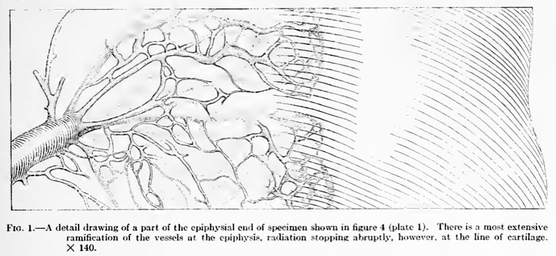
Fig. 1. Pigeon Bone Epiphysial End
A detail drawing of a part of the epiphysial end of specimen shown in figure 4 (plate 1). There is a most extensive ramification of the vessels at the epiphysis, radiation stopping abruptly, however, at the line of cartilage. X 140.
| Historic Disclaimer - information about historic embryology pages |
|---|
 |
File history
Click on a date/time to view the file as it appeared at that time.
| Date/Time | Thumbnail | Dimensions | User | Comment | |
|---|---|---|---|---|---|
| current | 15:38, 29 March 2011 | | 800 × 369 (79 KB) | S8600021 (talk | contribs) | ==Fig. 1. Pigeon Bone Epiphysial End== A detail drawing of a part of the epiphysial end of specimen shown in figure 4 (plate 1). There is a most extensive ramification of the vessels at the epiphysis, radiation stopping abruptly, however, at the line of |
You cannot overwrite this file.
File usage
The following page uses this file: