File:Dickie1914 fig03.jpg: Difference between revisions
mNo edit summary |
mNo edit summary |
||
| Line 1: | Line 1: | ||
==Fig. 3. Lateral | ==Fig. 3. Lateral Surface Model of Brain and Cranial Nerves== | ||
The primitive forebrain is distinctly separated into telencephalic and diencephalic portions. The telencephalon has divided into two cerebral hemispheres, and each hemisphere, which is ovoid in form, has grown ventrally (forwards) beyond the lamina terminalis and dorsally (backwards) over the antero-superior portion of the diencephalon (fig. 3). | |||
{{Dickie1914 figures}} | {{Dickie1914 figures}} | ||
Latest revision as of 22:14, 19 August 2015
Fig. 3. Lateral Surface Model of Brain and Cranial Nerves
The primitive forebrain is distinctly separated into telencephalic and diencephalic portions. The telencephalon has divided into two cerebral hemispheres, and each hemisphere, which is ovoid in form, has grown ventrally (forwards) beyond the lamina terminalis and dorsally (backwards) over the antero-superior portion of the diencephalon (fig. 3).
| Historic Disclaimer - information about historic embryology pages |
|---|
 |
Reference
Dickie JK. The anatomy of the head end of a 20 mm human embryo. (1914) J Anat Physiol., 48(4): 445-60. PMID 17233010
Cite this page: Hill, M.A. (2024, May 20) Embryology Dickie1914 fig03.jpg. Retrieved from https://embryology.med.unsw.edu.au/embryology/index.php/File:Dickie1914_fig03.jpg
- © Dr Mark Hill 2024, UNSW Embryology ISBN: 978 0 7334 2609 4 - UNSW CRICOS Provider Code No. 00098G
File history
Click on a date/time to view the file as it appeared at that time.
| Date/Time | Thumbnail | Dimensions | User | Comment | |
|---|---|---|---|---|---|
| current | 21:49, 19 August 2015 | 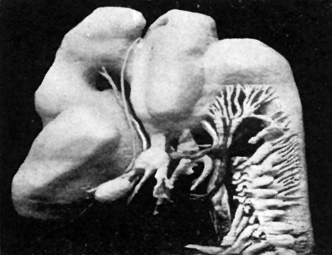 | 672 × 516 (64 KB) | Z8600021 (talk | contribs) | |
| 21:48, 19 August 2015 |  | 885 × 594 (83 KB) | Z8600021 (talk | contribs) | {{Dickie1914 figures}} |
You cannot overwrite this file.
File usage
The following 2 pages use this file: