File:DibleWest1941 fig03.jpg: Difference between revisions
From Embryology
No edit summary |
mNo edit summary |
||
| Line 1: | Line 1: | ||
==Fig. 3. Coloured drawing of a section (slide 25-2) through the middle of the ovum== | |||
x100. | |||
{{DibleWest1941 figures}} | |||
Revision as of 16:38, 28 July 2015
Fig. 3. Coloured drawing of a section (slide 25-2) through the middle of the ovum
x100.
| Historic Disclaimer - information about historic embryology pages |
|---|
 |
- Links: Fig. 1 | Fig. 2 | Fig. 3 | Fig. 4 | Fig. 5 | Fig. 6 | Fig. 7 | Fig. 8 | Fig. 9 | Fig. 10 | Fig. 11 | Plate 1 | Plate 2 | Plate 3 | Plate 4 | Plate 5 | Plate 6
Reference
Dible JH. and West CM. A human ovum at the previllous stage. (1941) J Anat. 75(3): 269–281. PMID 17104860
Cite this page: Hill, M.A. (2024, May 23) Embryology DibleWest1941 fig03.jpg. Retrieved from https://embryology.med.unsw.edu.au/embryology/index.php/File:DibleWest1941_fig03.jpg
- © Dr Mark Hill 2024, UNSW Embryology ISBN: 978 0 7334 2609 4 - UNSW CRICOS Provider Code No. 00098G
File history
Click on a date/time to view the file as it appeared at that time.
| Date/Time | Thumbnail | Dimensions | User | Comment | |
|---|---|---|---|---|---|
| current | 16:33, 28 July 2015 | 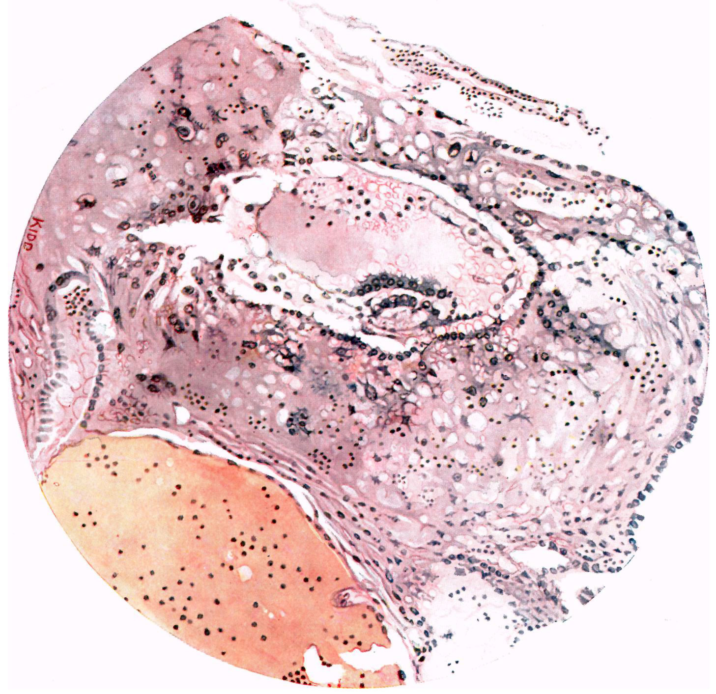 | 1,457 × 1,413 (458 KB) | Z8600021 (talk | contribs) |
You cannot overwrite this file.
File usage
The following page uses this file: