File:Cullen1916 fig31.jpg

Original file (1,280 × 1,072 pixels, file size: 355 KB, MIME type: image/jpeg)
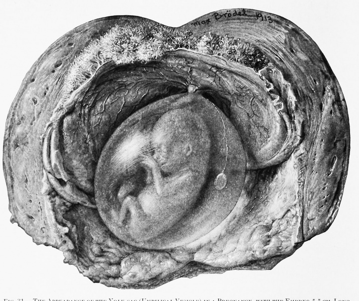
Fig. 31. The Appearance of the Yolk-sac (Umbilical Vesicle) in a Pregnancy with the Embryo 5.5 cm Long
The chorion has been split and turned back on the uterine wall. The amniotic cavity is still intact, appearing as a tensely filled oval cyst containing the embryo. Coming from the placental attachment of the umbilical cord is a delicate vaseular thread which passes to the flattened, yellowish, opaque yolk-sac, now referred to as the umbilical vesicle. It remains approximately this size until birth. (See Fig. 32.)
| Historic Disclaimer - information about historic embryology pages |
|---|
 |
- Figure Links: 1 Human embryo 0.7 mm | 2 Human embryo 1.7 mm | 3 Human embryo 2.5 mm | 4 Human embryo 3.5 mm | 5 Human embryo 5 mm | 6 Human embryo 7 mm | 7 Human embryo 7 mm | 8 Human embryo 10 mm | 9 Human embryo 12.5 mm | 10 Human embryo 10 mm | 11 Human embryo 23 mm | 12 Human embryo 3 cm | 13 Human embryo 4.5 cm sagittal | 14 Human Embryo 4.5 cm | 15 Human Embryo 5.2 cm | 16 Human Embryo 6.5 cm | 17 Human Embryo 7.5 cm | 18 Human Embryo 9 cm | 19 Human Embryo 10 cm | 20 Human Embryo 12 cm | 21 Human Embryo 12 cm | 22 Human Embryo 12 cm | 23 Human Embryo 12 cm Cord | 28 Fetus Five Months | 30 Ventral Heria | 31 Human Embryo 5.5 cm | 32 Term Human | 33 Term Human | [[Figures
Reference
Cullen TS. Embryology, anatomy, and diseases of the umbilicus together with diseases of the urachus. (1916) W. B. Saunders Company, Philadelphia And London.
Cite this page: Hill, M.A. (2024, April 28) Embryology Cullen1916 fig31.jpg. Retrieved from https://embryology.med.unsw.edu.au/embryology/index.php/File:Cullen1916_fig31.jpg
- © Dr Mark Hill 2024, UNSW Embryology ISBN: 978 0 7334 2609 4 - UNSW CRICOS Provider Code No. 00098G
File history
Click on a date/time to view the file as it appeared at that time.
| Date/Time | Thumbnail | Dimensions | User | Comment | |
|---|---|---|---|---|---|
| current | 14:00, 28 October 2018 | | 1,280 × 1,072 (355 KB) | Z8600021 (talk | contribs) | |
| 13:59, 28 October 2018 |  | 2,068 × 1,991 (957 KB) | Z8600021 (talk | contribs) |
You cannot overwrite this file.
File usage
The following 2 pages use this file: